What is myxoid stroma? Imagine a hidden world within your body, a delicate network of cells and molecules that provides structure and support for tissues. Myxoid stroma, a specialized type of connective tissue, is like the scaffolding that holds everything together, allowing cells to migrate, proliferate, and communicate.
This intricate structure, composed of a gel-like matrix and a variety of cells, plays a vital role in maintaining tissue integrity and facilitating essential biological processes. From the cartilage in your joints to the delicate membranes surrounding organs, myxoid stroma is everywhere, silently contributing to your body’s health.
Definition of Myxoid Stroma
Myxoid stroma is a type of connective tissue found in various biological tissues. It’s characterized by its jelly-like, mucoid appearance, which is a result of its unique composition.
Characteristics of Myxoid Stroma
Myxoid stroma is characterized by its distinctive appearance, composition, and function.
Appearance
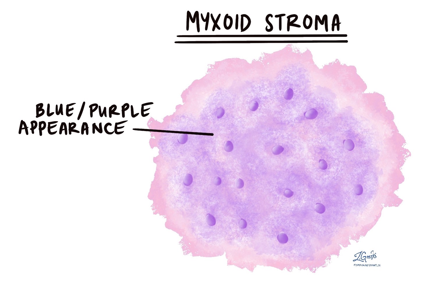
Myxoid stroma is typically translucent or slightly opaque, with a gelatinous or mucoid consistency. It often appears as a pale, homogeneous matrix under a microscope.
Composition
Myxoid stroma is primarily composed of a ground substance rich in hyaluronic acid, a type of glycosaminoglycan (GAG). Hyaluronic acid is a large, negatively charged molecule that attracts water, giving the stroma its characteristic gelatinous nature.
Function
Myxoid stroma serves several important functions:* Structural Support: It provides a framework for surrounding cells, supporting their organization and providing structural integrity to the tissue.
Diffusion
The gelatinous nature of myxoid stroma facilitates the diffusion of nutrients and oxygen to cells, as well as the removal of waste products.
Cell Migration
Myxoid stroma plays a role in cell migration, allowing cells to move through the tissue. This is particularly important during development and wound healing.
Examples of Tissues with Myxoid Stroma
Myxoid stroma is found in a variety of tissues, including:* Connective Tissues: It is a common component of loose connective tissue, where it provides support and flexibility.
Tumors
Myxoid stroma is often associated with tumors, particularly those of mesenchymal origin, such as liposarcoma.
Cartilage
It is present in the extracellular matrix of cartilage, contributing to its resilience and shock-absorbing properties.
Wharton’s Jelly
This is a specialized type of myxoid stroma found in the umbilical cord, providing support and protection to the developing fetus.
Brain
Myxoid stroma is also present in the brain, where it helps to maintain the structure of the nervous tissue.
Cellular Components of Myxoid Stroma: What Is Myxoid Stroma
Myxoid stroma, a specialized connective tissue characterized by its gelatinous appearance, is a vital component of various tissues and organs. It’s not just a passive structural element; it actively participates in tissue function and development. This intricate matrix is composed of a diverse array of cells, each contributing to the unique properties and functions of myxoid stroma.
Types of Cells in Myxoid Stroma
The cellular inhabitants of myxoid stroma are crucial to its structure and function. These cells, with their distinct morphologies and roles, work in concert to create a dynamic and responsive environment.
- Fibroblasts: These are the most abundant cells in myxoid stroma. They are responsible for synthesizing and maintaining the extracellular matrix (ECM), which is the gel-like substance that gives myxoid stroma its characteristic appearance. Fibroblasts produce collagen, hyaluronic acid, and other components that contribute to the ECM’s unique properties. They also play a role in wound healing and tissue repair.
- Myofibroblasts: These specialized fibroblasts exhibit features of both smooth muscle cells and fibroblasts. They possess contractile properties, allowing them to contribute to tissue contraction and remodeling. Myofibroblasts are particularly important in wound healing, where they help to close wounds and restore tissue integrity.
- Mast Cells: These cells are known for their role in immune responses and inflammation. They contain granules filled with histamine and other mediators that are released upon activation. Mast cells contribute to the inflammatory response in myxoid stroma, which can be important in wound healing and defense against pathogens.
- Macrophages: These phagocytic cells are responsible for clearing cellular debris, pathogens, and other foreign substances from myxoid stroma. They play a crucial role in maintaining tissue homeostasis and preventing infections. Macrophages also participate in immune responses and tissue repair.
- Pericytes: These cells wrap around blood vessels in myxoid stroma and contribute to vascular stability and regulation. They can differentiate into other cell types, including fibroblasts and smooth muscle cells, depending on the tissue’s needs. Pericytes are also involved in angiogenesis, the formation of new blood vessels.
Extracellular Matrix of Myxoid Stroma

The extracellular matrix (ECM) in myxoid stroma is a unique and specialized structure that plays a crucial role in the development, function, and behavior of the surrounding tissues. It’s like a complex scaffolding system, providing structural support, influencing cell communication, and facilitating tissue growth and repair.
Composition of the Extracellular Matrix
The ECM of myxoid stroma is primarily composed of a gel-like substance rich in water, glycosaminoglycans (GAGs), and proteoglycans. This composition gives the ECM its characteristic myxoid appearance, meaning it’s soft and jelly-like.
Role of Specific Components
- Hyaluronic Acid: This unbranched GAG is a major component of the myxoid stroma ECM. It’s a highly hydrated molecule that attracts and retains water, contributing to the gel-like consistency of the ECM. Hyaluronic acid acts as a space-filling molecule, creating a hydrated environment that allows for cell migration and diffusion of nutrients and waste products.
- Proteoglycans: These are complex molecules composed of a core protein attached to GAG chains. Proteoglycans are abundant in myxoid stroma and contribute to its structural integrity and its ability to bind to growth factors and other signaling molecules.
- Aggrecan: A large proteoglycan found in myxoid stroma, it interacts with hyaluronic acid to form large aggregates, further contributing to the gel-like nature of the ECM.
- Versican: Another important proteoglycan, it’s involved in cell adhesion and migration, and it can also regulate the activity of growth factors.
- Collagen: Although less abundant than GAGs and proteoglycans, collagen is present in myxoid stroma and provides structural support. The type of collagen present can vary depending on the specific tissue and its developmental stage.
- Type I collagen: Provides tensile strength and contributes to the overall structure of the ECM.
- Type III collagen: More flexible than type I collagen and plays a role in tissue repair and regeneration.
Summary of Extracellular Matrix Components and Functions
| Component | Function |
|---|---|
| Hyaluronic Acid | Hydration, space-filling, cell migration, diffusion of molecules |
| Proteoglycans (Aggrecan, Versican) | Structural integrity, binding to growth factors and signaling molecules, cell adhesion and migration |
| Collagen (Type I, Type III) | Structural support, tensile strength, tissue repair and regeneration |
Physiological Significance of Myxoid Stroma
The myxoid stroma, with its unique composition of cells and extracellular matrix, plays a crucial role in supporting and maintaining the structural integrity of various tissues and organs. It provides a dynamic environment that facilitates essential cellular processes like migration, proliferation, and differentiation, ultimately contributing to the overall functionality of the tissues it supports.
Role in Tissue Support and Integrity
The myxoid stroma acts as a scaffold, providing structural support to the surrounding cells and tissues. The gel-like consistency of the extracellular matrix provides a cushioning effect, protecting cells from mechanical stress and damage. The presence of collagen fibers within the stroma further enhances its tensile strength, enabling it to withstand stretching and compression forces. This structural support is particularly important in tissues that are subjected to constant movement or pressure, such as muscles, tendons, and ligaments.
Influence on Cell Migration, Proliferation, and Differentiation, What is myxoid stroma
The myxoid stroma actively participates in regulating cellular processes by providing a dynamic environment that influences cell behavior. The composition of the extracellular matrix, particularly the presence of growth factors and signaling molecules, plays a crucial role in guiding cell migration and proliferation. The stroma acts as a conduit for these signaling molecules, ensuring their delivery to the target cells, thereby controlling their movement and division.
Furthermore, the stroma influences cell differentiation by providing cues that direct cells to adopt specific fates and functions. This intricate interplay between the stroma and cells is essential for tissue development, repair, and regeneration.
Examples of Myxoid Stroma Function in Specific Tissues
The myxoid stroma plays a vital role in the function of various organs and tissues. For example, in the brain, the myxoid stroma, known as the perineuronal net, surrounds neurons and regulates their activity. The net provides structural support to the neurons and influences their synaptic plasticity, contributing to learning and memory formation. In the gastrointestinal tract, the myxoid stroma in the lamina propria supports the epithelial lining and contributes to nutrient absorption and immune function.
In the musculoskeletal system, the myxoid stroma within tendons and ligaments facilitates their flexibility and strength, allowing for smooth movement and joint stability.
Myxoid Stroma in Disease

Myxoid stroma, while normally a supportive component of tissues, can become altered in various diseases, including cancer. These alterations can contribute to the development and progression of disease, making the myxoid stroma a potential target for therapeutic interventions.
Association of Myxoid Stroma with Diseases
The presence of myxoid stroma can be associated with a range of diseases, including:
- Cancer: Myxoid stroma is commonly found in various types of cancer, including:
- Liposarcoma: A type of soft tissue sarcoma characterized by the presence of abundant myxoid stroma.
- Myxoid chondrosarcoma: A type of cartilage cancer that often exhibits a myxoid matrix.
- Breast cancer: Myxoid stroma can be present in some breast cancers and has been linked to increased tumor growth and metastasis.
- Fibromuscular dysplasia (FMD): A condition affecting the blood vessels, where myxoid stroma can contribute to the thickening and narrowing of the vessel walls.
- Inflammatory conditions: Myxoid stroma can be present in inflammatory conditions like rheumatoid arthritis, where it contributes to joint swelling and inflammation.
Alterations in Myxoid Stroma and Disease Progression
Alterations in the composition and structure of myxoid stroma can contribute to disease development and progression in several ways:
- Increased tumor growth and metastasis: In cancer, myxoid stroma can provide a favorable environment for tumor growth and metastasis by:
- Providing a scaffold for tumor cells: Myxoid stroma can provide a network of support for tumor cells, allowing them to grow and spread more easily.
- Secreting growth factors: Myxoid stroma can produce growth factors that stimulate tumor cell proliferation and angiogenesis (formation of new blood vessels), which further supports tumor growth.
- Promoting tumor cell invasion: Myxoid stroma can create a looser, more permeable environment, facilitating the invasion of tumor cells into surrounding tissues.
- Impaired tissue function: In conditions like FMD, the altered myxoid stroma can lead to thickening and narrowing of blood vessels, impairing blood flow and causing symptoms like hypertension and stroke.
- Increased inflammation: In inflammatory conditions, the presence of myxoid stroma can contribute to chronic inflammation by attracting inflammatory cells and releasing pro-inflammatory mediators.
Therapeutic Implications of Targeting Myxoid Stroma
The role of myxoid stroma in disease progression suggests that targeting this component could offer therapeutic benefits. Several approaches are being explored:
- Anti-angiogenic therapies: Inhibiting the formation of new blood vessels in tumors by targeting factors produced by myxoid stroma could reduce tumor growth and metastasis.
- Targeting stromal-derived growth factors: Blocking the action of growth factors produced by myxoid stroma could inhibit tumor cell proliferation and angiogenesis.
- Modulating the composition of myxoid stroma: Modifying the composition of myxoid stroma to make it less supportive of tumor growth or more conducive to tissue repair could be a promising therapeutic strategy.
Methods for Studying Myxoid Stroma

Understanding the structure and function of myxoid stroma requires a multifaceted approach, utilizing various techniques to delve into its intricate components. These methods provide valuable insights into the roles of myxoid stroma in normal physiology and disease pathogenesis.
Histological Analysis
Histological analysis is a cornerstone of myxoid stroma research, providing a visual representation of its structural organization. This technique involves preparing thin sections of tissue, staining them with specific dyes, and examining them under a microscope.
- Hematoxylin and eosin (H&E) staining is a routine staining method that highlights the nuclei (blue) and cytoplasm (pink) of cells, allowing for visualization of cellular morphology and arrangement.
- Periodic acid-Schiff (PAS) staining specifically stains carbohydrates, including the abundant glycosaminoglycans present in the extracellular matrix of myxoid stroma. This method helps to visualize the distribution and density of these important components.
- Alcian blue staining is another technique that highlights acidic glycosaminoglycans, further enhancing the understanding of the composition and organization of the extracellular matrix.
Histological analysis provides valuable information about the overall structure of myxoid stroma, including the distribution and morphology of its cellular components and the nature of its extracellular matrix. However, this technique is limited in its ability to provide detailed information about the specific molecules and their interactions within the stroma.
Immunohistochemistry
Immunohistochemistry (IHC) is a powerful technique that uses antibodies to specifically detect and localize target molecules within tissue sections. This method allows for the identification and characterization of various proteins, including those involved in cell signaling, adhesion, and matrix remodeling.
- Antibodies targeting specific cell markers, such as vimentin, CD34, and S100, can be used to identify and quantify the different cell types present in myxoid stroma.
- Antibodies against extracellular matrix components, such as collagen, laminin, and fibronectin, can be used to assess the composition and organization of the matrix.
- Antibodies targeting signaling molecules, such as growth factors and their receptors, can be used to study the role of myxoid stroma in cell proliferation, differentiation, and migration.
IHC provides valuable information about the specific molecules and their interactions within myxoid stroma, but it is limited by the availability of specific antibodies and the potential for cross-reactivity.
Molecular Biology Techniques
Molecular biology techniques provide a deeper understanding of the molecular mechanisms underlying myxoid stroma function. These techniques allow for the analysis of gene expression, protein interactions, and signaling pathways.
- Reverse transcription polymerase chain reaction (RT-PCR) is a technique used to measure the expression levels of specific genes in myxoid stroma. This method can be used to identify genes that are differentially expressed in diseased versus normal stroma.
- Microarray analysis allows for the simultaneous measurement of the expression levels of thousands of genes. This technique can be used to identify gene expression signatures associated with specific myxoid stroma phenotypes.
- Next-generation sequencing (NGS) is a high-throughput sequencing technique that can be used to analyze the complete genome or transcriptome of myxoid stroma cells. This technique can be used to identify genetic mutations and gene expression changes that contribute to disease pathogenesis.
- Chromatin immunoprecipitation (ChIP) is a technique used to identify the proteins that bind to specific regions of DNA. This method can be used to study the regulation of gene expression in myxoid stroma cells.
Molecular biology techniques provide a comprehensive understanding of the molecular mechanisms underlying myxoid stroma function, but they can be complex and require specialized equipment and expertise.
Table Summarizing Methods and Applications
| Method | Application | Advantages | Limitations |
|---|---|---|---|
| Histological Analysis | Visualize structural organization, identify cell types, assess matrix composition | Relatively simple and inexpensive | Limited molecular specificity, requires skilled interpretation |
| Immunohistochemistry | Identify and localize specific molecules, study protein interactions | High specificity, can be used on tissue sections | Availability of specific antibodies, potential for cross-reactivity |
| Molecular Biology Techniques | Analyze gene expression, protein interactions, signaling pathways | Comprehensive understanding of molecular mechanisms | Complex and expensive, requires specialized equipment and expertise |
Understanding myxoid stroma opens a window into the complex interplay of cells and their environment. Its role in tissue development, function, and even disease progression makes it a fascinating area of study. Whether you’re a biology enthusiast or simply curious about the inner workings of your body, myxoid stroma offers a glimpse into the hidden world that keeps you going.
Frequently Asked Questions
What are some examples of tissues where myxoid stroma is found?
Myxoid stroma is found in various tissues, including cartilage, tendons, ligaments, and some types of tumors. It’s also present in the stroma of organs like the heart, lungs, and kidneys.
How does myxoid stroma contribute to the development of disease?
Alterations in myxoid stroma composition and structure can contribute to the development and progression of various diseases, including cancer. For example, in some cancers, the myxoid stroma can become abnormally dense, promoting tumor growth and invasion.
What are some potential therapeutic implications of targeting myxoid stroma?
Targeting myxoid stroma in disease treatment is a promising area of research. By manipulating the composition or function of myxoid stroma, scientists hope to develop new therapies to control tumor growth, enhance tissue regeneration, and improve treatment outcomes.


