What is the stroma of the eye? It’s the supportive framework that keeps your eye strong and healthy. Imagine it like the scaffolding of a building, providing structure and holding everything together. The stroma is made up of strong fibers, like collagen and elastin, and a gel-like substance that cushions and nourishes the eye. It’s also responsible for how light travels through your eye, allowing you to see clearly.
The stroma is a crucial part of the cornea and sclera, the transparent front part and the white outer layer of the eye respectively. These layers are responsible for maintaining the eye’s shape, protecting the delicate inner structures, and allowing light to pass through. Understanding the stroma is key to appreciating the complexity of the eye and the delicate balance that makes vision possible.
What is the Stroma?
The stroma is a crucial component of the eye, acting as the supportive framework for the cornea and sclera, the two outer layers of the eye. It provides structural integrity and plays a vital role in maintaining the eye’s shape and function.
Stroma Composition, What is the stroma of the eye
The stroma is primarily composed of a dense network of collagen fibers, elastin fibers, and ground substance.
- Collagen fibers are the most abundant component, providing tensile strength and rigidity to the stroma. These fibers are arranged in a highly organized, layered structure, contributing to the cornea’s transparency.
- Elastin fibers provide flexibility and elasticity, allowing the stroma to stretch and return to its original shape. This property is essential for maintaining the eye’s shape and accommodating changes in pressure.
- Ground substance is a gel-like matrix that fills the spaces between the collagen and elastin fibers. It is composed of water, proteoglycans, and glycoproteins, providing hydration and lubrication to the stroma.
Stroma Connection to Cornea and Sclera
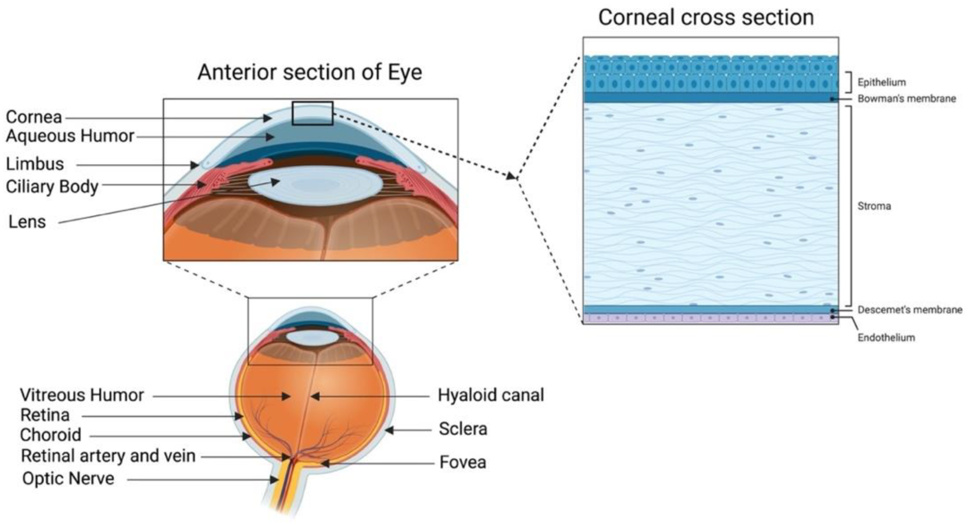
The stroma forms the bulk of the cornea, the transparent outer layer of the eye responsible for focusing light. The stroma’s highly organized structure, with its parallel arrangement of collagen fibers, ensures that light passes through the cornea with minimal scattering, contributing to clear vision. The stroma also constitutes the middle layer of the sclera, the white outer layer of the eye.
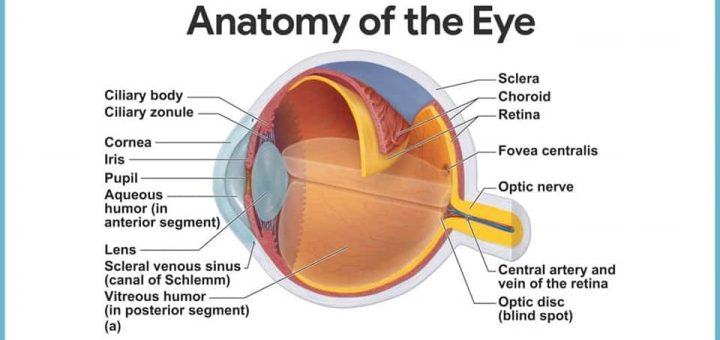
The sclera provides structural support and protection to the eye. The stroma in the sclera is less organized than in the cornea, containing a higher proportion of elastin fibers, which allows the sclera to maintain its shape while accommodating changes in pressure.
Location and Structure of the Stroma

The stroma is the main structural component of the cornea, the transparent outer layer of the eye that plays a crucial role in focusing light onto the retina. It is a dense, fibrous, and avascular (lacking blood vessels) tissue that provides the cornea with its strength and rigidity. Understanding the location and structure of the stroma is essential for comprehending how the cornea functions effectively.
Stroma’s Location Within the Eye
The stroma is sandwiched between two other layers of the cornea: the epithelium on the outer surface and the endothelium on the inner surface. It accounts for approximately 90% of the cornea’s total thickness.
- Epithelium: This outermost layer is a thin, transparent sheet of cells that protects the cornea from the external environment and aids in maintaining its hydration.
- Stroma: The middle layer, the stroma, is the thickest and most substantial layer of the cornea. It provides the cornea with its structural integrity and transparency.
- Endothelium: This innermost layer is a single layer of cells that helps regulate the flow of fluids in and out of the cornea, maintaining its hydration and transparency.
Layered Structure of the Stroma
The stroma is composed of numerous layers of collagen fibrils, which are thin, elongated protein fibers that are arranged in a highly organized, parallel fashion. These fibrils are embedded in a matrix of proteoglycans, which are complex sugar molecules that help to hydrate and maintain the spacing between the collagen fibrils.
- The collagen fibrils in each layer are aligned parallel to each other, but the orientation of the fibrils in adjacent layers is slightly different. This staggered arrangement of collagen fibrils contributes to the cornea’s strength and ability to withstand pressure.
- The highly organized structure of the stroma is essential for maintaining the cornea’s transparency. The precise spacing between the collagen fibrils and the presence of proteoglycans allow light to pass through the cornea without being scattered, ensuring clear vision.
Layers of the Stroma
The stroma is typically divided into several distinct layers based on the size and organization of the collagen fibrils:
| Layer | Characteristics |
|---|---|
| Anterior Stroma | – Contains smaller, more densely packed collagen fibrils.
|
| Mid-Stroma | – Contains larger, more loosely packed collagen fibrils.
|
| Posterior Stroma | – Contains the largest collagen fibrils.
|
Functions of the Stroma
The stroma, the dense connective tissue of the eye, plays a crucial role in maintaining the eye’s structure and function. It acts as the supporting framework, providing a platform for other ocular tissues and contributing to the overall integrity of the eye.
Role in Maintaining the Eye’s Shape and Integrity
The stroma provides structural support to the cornea, the transparent outer layer of the eye, which is responsible for focusing light onto the retina. The stroma’s collagen fibers are arranged in a highly organized, layered pattern, providing strength and rigidity to the cornea. This organized structure allows the cornea to maintain its shape and resist deformation, which is essential for clear vision.
Role in Light Transmission and Refraction
The stroma’s unique structure, characterized by its high water content and precisely arranged collagen fibers, plays a vital role in light transmission and refraction. The collagen fibers, being transparent, allow light to pass through the cornea with minimal scattering. The stroma’s regular arrangement of collagen fibers ensures that light is refracted (bent) in a predictable manner, contributing to the eye’s focusing ability.
Role in Nutrient and Waste Exchange
The stroma acts as a conduit for nutrient and waste exchange within the cornea. The stroma’s vascular network, located in the limbus (the outer edge of the cornea), supplies oxygen and nutrients to the avascular corneal cells. The stroma also facilitates the removal of waste products from the corneal cells, maintaining a healthy corneal environment.
Stroma and Vision: What Is The Stroma Of The Eye

The stroma, as the structural backbone of the cornea, plays a crucial role in maintaining the clarity and shape of the eye, directly influencing visual acuity. Any abnormalities in the stroma can lead to vision problems, impacting the ability to see clearly.
Impact of Stromal Abnormalities on Vision
Stromal abnormalities can disrupt the cornea’s transparency and curvature, leading to a range of vision issues. The most common consequences include:
- Reduced Visual Acuity: Irregularities in the stroma’s structure can scatter light, blurring vision and making it difficult to see fine details.
- Astigmatism: An uneven curvature of the cornea, often caused by stromal abnormalities, results in distorted vision, particularly at different angles.
- Myopia (Nearsightedness): A thicker or more curved stroma can cause light to focus in front of the retina, making distant objects appear blurry.
- Hyperopia (Farsightedness): A thinner or less curved stroma can cause light to focus behind the retina, making near objects appear blurry.
Common Conditions Affecting the Stroma
Several conditions can directly affect the stroma, leading to impaired vision. Two prominent examples include:
- Keratoconus: This condition involves a progressive thinning and bulging of the cornea, resulting in a cone-shaped distortion. Keratoconus causes blurred vision, double vision, and sensitivity to light.
- Corneal Dystrophies: These are genetic disorders that affect the cornea’s structure, leading to abnormal deposits in the stroma. Corneal dystrophies can cause clouding of the cornea, impairing vision.
Role of the Stroma in Refractive Surgery Procedures
The stroma plays a central role in refractive surgery procedures, such as LASIK and PRK, which aim to correct refractive errors by reshaping the cornea. During these procedures, a flap of corneal tissue is created, and the stroma is precisely reshaped using a laser. The stromal modifications alter the cornea’s curvature, changing how light focuses on the retina, improving vision.
Research and Future Directions

The stroma is a dynamic and complex structure that continues to be a subject of intense scientific investigation. Recent research advancements are shedding new light on its intricate roles in eye health and disease. This understanding has opened up exciting avenues for potential therapeutic interventions and novel approaches to ophthalmic care.
Stromal Research Advancements
Recent research has made significant strides in understanding the stromal microenvironment and its influence on ocular health. For example, studies have explored the role of stromal cells in wound healing and regeneration.
- Researchers have identified specific stromal cell populations, such as keratocytes and fibroblasts, that contribute to corneal wound repair. These cells are responsible for producing the extracellular matrix components that are essential for tissue regeneration.
- Studies have also investigated the role of stromal cells in the immune response of the eye. Stromal cells can interact with immune cells, such as lymphocytes and macrophages, to regulate inflammation and protect against pathogens.
Furthermore, investigations into the molecular mechanisms underlying stromal function have yielded promising insights.
- Studies have identified various signaling pathways and growth factors that regulate stromal cell behavior and contribute to the maintenance of corneal transparency. These findings could potentially lead to the development of new therapies for corneal diseases.
- Researchers have also explored the role of microRNAs in stromal function. MicroRNAs are small, non-coding RNA molecules that regulate gene expression. Studies have shown that specific microRNAs are involved in stromal cell differentiation, proliferation, and wound healing.
The stroma is a remarkable structure that plays a vital role in the intricate workings of the eye. Its ability to provide support, regulate light transmission, and facilitate nutrient exchange highlights its importance in maintaining healthy vision. While the stroma might not be the most visible part of the eye, its influence is profound. It’s a testament to the incredible complexity and elegance of the human body, and its significance in allowing us to experience the world around us through the gift of sight.
Question & Answer Hub
What happens if the stroma is damaged?
Damage to the stroma can lead to a variety of vision problems, depending on the severity and location of the damage. Some common conditions affecting the stroma include keratoconus, corneal dystrophies, and scarring. These conditions can cause blurred vision, distortion, and even blindness if left untreated.
Can the stroma be repaired?
Yes, there are various treatments available for stromal abnormalities, depending on the specific condition. These treatments can range from simple eye drops to more complex procedures like corneal transplants or refractive surgery.
What are some ongoing research areas related to the stroma?
Researchers are constantly exploring new ways to understand and treat stromal conditions. Some areas of focus include developing new therapies for keratoconus, improving corneal transplant techniques, and exploring the potential of stem cell therapy for corneal regeneration.


