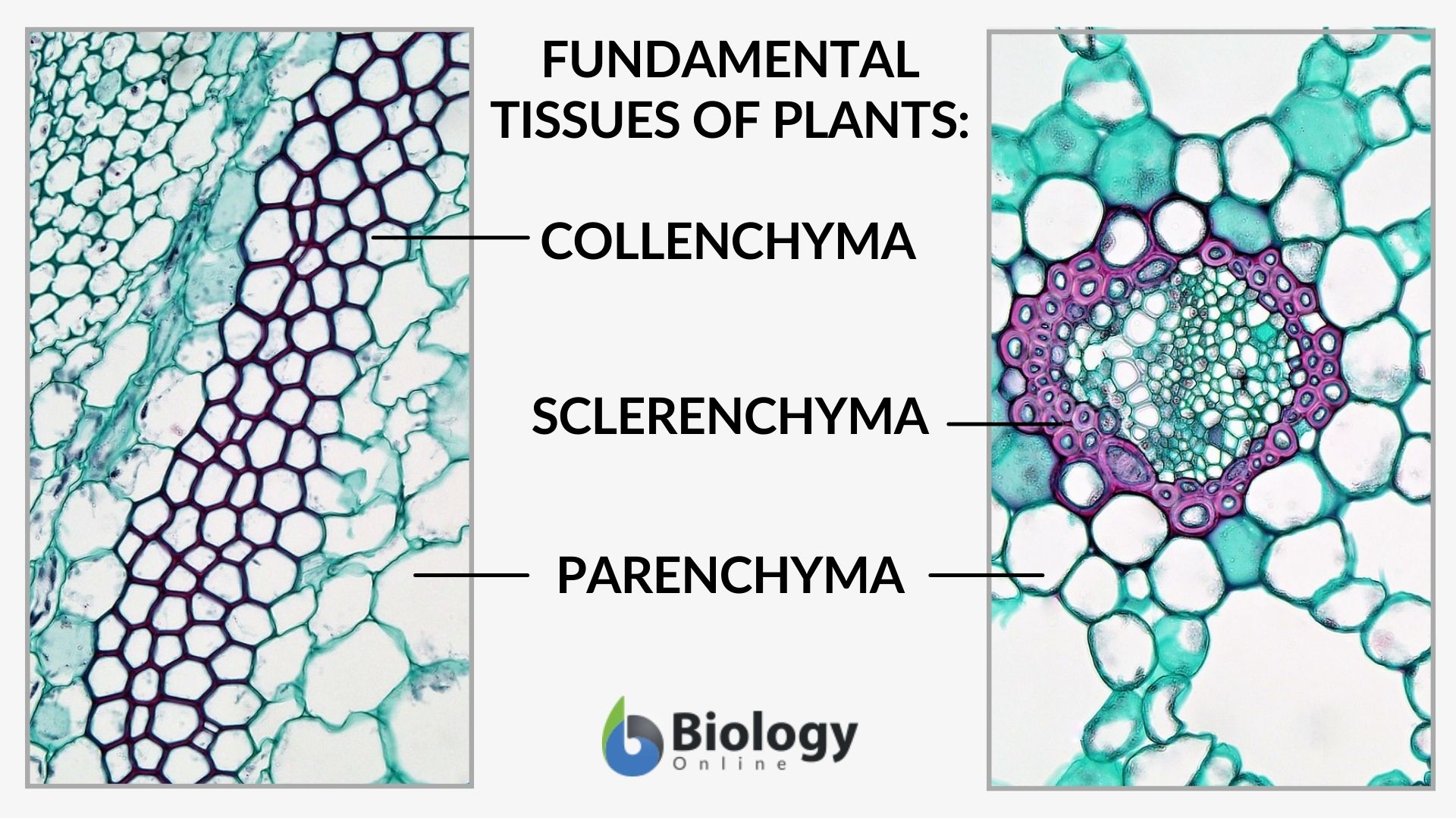
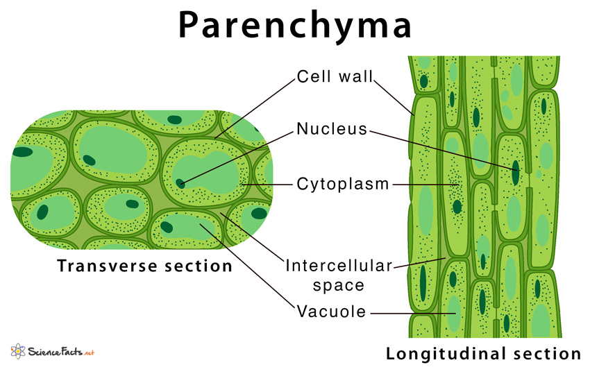
Are duct stroma or parenchyma – Are ducts stroma or parenchyma? This question delves into the fundamental organization of organs, revealing the intricate interplay between structural support and functional units. Ductal structures, found in various organs like salivary glands, pancreas, and mammary glands, serve as pathways for transporting essential substances. Understanding the distinction between stroma, the supporting framework, and parenchyma, the functional tissue, provides crucial insight into how organs operate.
Imagine a bustling city, where the roads, bridges, and utilities represent the stroma, providing the essential infrastructure. The buildings, businesses, and residents within the city represent the parenchyma, carrying out the city’s primary functions. Similarly, within organs, stroma and parenchyma work in harmony to maintain the organ’s integrity and facilitate its specific tasks.
Introduction to Ductal Structures: Are Duct Stroma Or Parenchyma
Ductal structures are a fundamental component of many organs, playing a crucial role in the transport of fluids and substances throughout the body. These structures are essentially tubular pathways that facilitate the movement of various materials, contributing to the overall function of the organ.
Examples of Ductal Structures
Ductal structures are found in various organs, each with a specific function. Here are some examples:
- Salivary Glands: Salivary glands, responsible for producing saliva, contain ducts that transport saliva from the glands to the oral cavity. The salivary ducts, such as the parotid duct, submandibular duct, and sublingual duct, carry saliva containing digestive enzymes and lubrication agents to the mouth.
- Pancreas: The pancreas, an organ involved in both digestion and hormone production, has ducts that carry pancreatic juice containing digestive enzymes to the small intestine. The main pancreatic duct, along with the common bile duct, empties into the duodenum, the first segment of the small intestine.
- Mammary Glands: Mammary glands, responsible for milk production, contain a complex network of ducts that transport milk from the milk-producing lobules to the nipple. These ducts branch extensively within the breast tissue, forming a system that efficiently delivers milk to the infant during lactation.
Function of Ducts in Transporting Substances
Ducts are essentially tubular structures that act as conduits for the transport of fluids and substances. They play a critical role in the movement of materials within and between organs, facilitating various physiological processes. The function of ducts can be summarized as follows:
Ducts provide a pathway for the efficient transport of fluids and substances from their source to their destination, ensuring the proper functioning of various organs and systems.
The specific substances transported by ducts vary depending on the organ and its function. For instance, salivary ducts carry saliva containing digestive enzymes and lubrication agents, while pancreatic ducts transport pancreatic juice rich in digestive enzymes. Mammary ducts transport milk from the milk-producing lobules to the nipple.The movement of substances through ducts is often driven by a combination of factors, including:
- Pressure gradients: Differences in pressure between the source and destination can drive the flow of fluids through ducts.
- Muscle contractions: Smooth muscle cells lining the walls of some ducts can contract, propelling substances forward.
- Ciliary action: In some cases, cilia, tiny hair-like structures, can beat rhythmically to move substances along the duct.
Understanding Stroma and Parenchyma
Organs are complex structures composed of different tissues that work together to perform specific functions. Within an organ, two key components contribute to its overall structure and function: stroma and parenchyma. Understanding the distinct roles of stroma and parenchyma is essential for comprehending the organization and operation of organs.
Stroma and its Supporting Role
Stroma refers to the connective tissue framework that provides support and structure to an organ. It acts as a scaffolding, holding together the functional cells and tissues of the organ. The stroma is composed of various components, including:
- Connective Tissue Fibers: Collagen, elastin, and reticular fibers provide strength, flexibility, and support to the organ. These fibers form a network that helps maintain the organ’s shape and integrity.
- Extracellular Matrix: This gel-like substance fills the spaces between cells and fibers, providing a medium for nutrient and waste exchange. It also helps regulate cell growth and differentiation.
- Blood Vessels: Stroma contains blood vessels that supply oxygen and nutrients to the parenchyma and remove waste products.
- Lymphatics: Lymphatic vessels within the stroma help drain excess fluid and transport immune cells.
The stroma is essential for maintaining the structural integrity of organs, allowing them to withstand mechanical stress and maintain their shape. It also provides a pathway for the diffusion of nutrients and oxygen to the parenchyma.
Parenchyma and its Functional Role
Parenchyma refers to the functional cells of an organ that perform its primary functions. These cells are specialized for specific tasks, such as secretion, absorption, filtration, or contraction. The parenchyma is responsible for the unique characteristics and activities of each organ. Examples of parenchyma include:
- Liver: Hepatocytes, the parenchymal cells of the liver, are responsible for detoxification, protein synthesis, and bile production.
- Kidney: Nephrons, the functional units of the kidney, are composed of parenchymal cells that filter blood and produce urine.
- Lungs: Alveolar epithelial cells, the parenchymal cells of the lungs, are responsible for gas exchange.
Interdependence of Stroma and Parenchyma, Are duct stroma or parenchyma
Stroma and parenchyma are interdependent and work together to ensure the proper functioning of an organ. The stroma provides a supportive framework for the parenchyma, allowing it to function effectively. For example, the blood vessels in the stroma deliver oxygen and nutrients to the parenchymal cells, while the lymphatic vessels remove waste products. In turn, the parenchyma produces substances that influence the stroma, such as growth factors that promote tissue repair and regeneration.
The relationship between stroma and parenchyma is analogous to a building’s foundation and walls. The foundation provides support and stability, while the walls are responsible for the building’s specific functions.
Ductal Stroma

The ductal stroma is the supportive framework that surrounds and interacts with the epithelial cells of ducts, providing structural integrity, facilitating communication, and regulating growth and development.
Components of Ductal Stroma
The ductal stroma is composed of various essential components that work together to maintain the integrity and functionality of the ducts.
- Connective Tissue: Connective tissue forms the primary structural component of the stroma, providing support and organization. It consists of various cells, including fibroblasts, myofibroblasts, and immune cells, embedded in an extracellular matrix composed of collagen, elastin, and proteoglycans. These components contribute to the tensile strength, elasticity, and resilience of the ductal stroma, enabling it to withstand mechanical stress and maintain its shape.
- Blood Vessels: A dense network of blood vessels permeates the ductal stroma, supplying oxygen and nutrients to the epithelial cells and removing waste products. The vascular network also plays a crucial role in regulating blood flow and pressure within the ducts, influencing the overall function and health of the epithelial cells.
- Nerves: Nerves innervate the ductal stroma, facilitating communication between the epithelial cells and the central nervous system. These nerves regulate the contraction and relaxation of smooth muscle cells within the stroma, influencing the flow of fluids through the ducts. They also play a role in sensing changes in the environment, such as pressure or inflammation, and transmitting signals to the nervous system.
- Lymphatics: Lymphatic vessels are essential for maintaining fluid balance and immune surveillance within the ductal stroma. They collect excess fluid and waste products from the epithelial cells and transport them to the lymph nodes, where immune cells can identify and eliminate pathogens. The lymphatic system also plays a role in the transport of immune cells and signaling molecules, contributing to the overall immune response within the ductal stroma.
Contribution to Ductal Integrity and Function
The various components of the ductal stroma work together to maintain the structural integrity and function of the ducts.
- Structural Support: Connective tissue provides a strong framework that supports the epithelial cells, preventing them from collapsing or distorting under pressure. This is particularly important in ducts that are subjected to significant mechanical stress, such as those in the urinary system or the gastrointestinal tract.
- Fluid Transport: The smooth muscle cells within the stroma, regulated by nerves, contract and relax to control the flow of fluids through the ducts. This ensures that fluids are transported efficiently and at the appropriate rate, preventing stagnation and facilitating the delivery of essential substances to the epithelial cells.
- Communication and Regulation: The nerves within the stroma allow for communication between the epithelial cells and the central nervous system, enabling the body to regulate the function of the ducts. This communication is essential for maintaining homeostasis and responding to changes in the environment.
- Immune Defense: The lymphatic vessels within the stroma transport immune cells and signaling molecules, facilitating the immune response to pathogens and inflammation. This helps to protect the epithelial cells from infection and maintain the health of the ducts.
Role in Ductal Growth and Development
The ductal stroma plays a crucial role in regulating the growth and development of ducts.
- Scaffolding for Growth: The connective tissue within the stroma provides a scaffold for the epithelial cells to grow and differentiate. This scaffolding ensures that the epithelial cells are organized properly and that the duct develops into the correct shape and size.
- Signaling Molecules: The stroma secretes various signaling molecules that influence the growth and differentiation of the epithelial cells. These molecules can stimulate or inhibit cell proliferation, migration, and differentiation, ensuring that the duct develops correctly and maintains its function.
- Vascular Supply: The vascular network within the stroma provides the epithelial cells with the necessary oxygen and nutrients to grow and develop. This is particularly important during periods of rapid growth, such as during embryonic development or tissue regeneration.
Ductal Parenchyma

The ductal parenchyma is the functional unit of the ductal system, responsible for the production, modification, and transport of various fluids, including saliva, sweat, and milk. It is composed of a specialized epithelium that forms the lining of the ducts, along with supporting cells called myoepithelial cells. These cells work together to facilitate the efficient transport and secretion of fluids.
Cell Types in Ductal Parenchyma
The ductal parenchyma is comprised of three main cell types: epithelial cells, myoepithelial cells, and secretory cells. Each cell type plays a distinct role in the overall function of the ductal system.
- Epithelial Cells: These cells form the inner lining of the ducts and are responsible for regulating the movement of fluids and solutes between the lumen of the duct and the surrounding tissue. Epithelial cells can be classified into different types based on their structure and function, such as squamous, cuboidal, and columnar epithelium.
- Myoepithelial Cells: These cells are located beneath the epithelial cells and are characterized by their contractile properties.
Myoepithelial cells play a crucial role in propelling fluids along the ducts by contracting and squeezing the ductal lumen. This contractile force is essential for the efficient movement of secretions and waste products.
- Secretory Cells: These cells are responsible for the production and release of specific substances, such as enzymes, hormones, or other specialized molecules. Secretory cells are often found in specific regions of the ductal system, depending on the type of fluid being produced.
For example, in salivary glands, secretory cells are responsible for producing saliva, while in mammary glands, they produce milk.
Organization of Ductal Parenchyma
The organization of ductal parenchyma is designed to facilitate efficient transport and secretion. The ductal system is typically organized in a hierarchical manner, with smaller, more numerous terminal ducts merging into larger, fewer collecting ducts. This branching structure allows for the gradual modification and concentration of fluids as they move through the ductal system.
The efficient transport and secretion of fluids is crucial for maintaining homeostasis and performing essential physiological functions.
The epithelial cells lining the ducts are often specialized to perform specific functions, such as absorption, secretion, or filtration. The arrangement of epithelial cells can vary depending on the location and function of the duct. For example, in the salivary glands, the epithelial cells lining the terminal ducts are responsible for producing saliva, while those lining the collecting ducts are responsible for modifying and concentrating the saliva before it is released into the oral cavity.The myoepithelial cells are also strategically located to facilitate efficient fluid movement.
These cells are positioned between the epithelial cells and the basement membrane, allowing them to exert a contractile force on the ductal lumen. The coordinated contraction of myoepithelial cells along the ductal system helps to propel fluids in the desired direction.The organization of ductal parenchyma, with its specialized cells and hierarchical structure, ensures the efficient transport and secretion of fluids, playing a vital role in maintaining homeostasis and supporting various physiological functions.
Examples of Ductal Stroma and Parenchyma

Understanding the specific components of ductal stroma and parenchyma is crucial for comprehending the functional organization of various organs. This section will delve into examples of different organs and their associated ductal structures, highlighting the unique characteristics of their stroma and parenchyma.
Examples of Ductal Stroma and Parenchyma in Different Organs
The following table provides a comprehensive overview of various organs and their associated ductal structures, outlining the key components of their stroma and parenchyma:
| Organ | Duct Type | Stroma Components | Parenchyma Components |
|---|---|---|---|
| Salivary Glands | Exocrine Ducts | Connective tissue, blood vessels, nerves | Acinar cells, ductal cells, myoepithelial cells |
| Pancreas | Exocrine Ducts | Connective tissue, blood vessels, nerves | Acinar cells, ductal cells, islet cells |
| Liver | Bile Ducts | Connective tissue, blood vessels, nerves | Hepatocytes, bile duct epithelial cells, Kupffer cells |
| Mammary Gland | Lactiferous Ducts | Connective tissue, adipose tissue, blood vessels, nerves | Epithelial cells of the ducts and alveoli, myoepithelial cells |
| Kidney | Collecting Ducts | Connective tissue, blood vessels, nerves | Epithelial cells of the tubules, glomerular cells |
Salivary Glands: The salivary glands are responsible for producing saliva, which plays a crucial role in digestion, lubrication, and oral hygiene. The parenchyma of salivary glands is composed of acinar cells, which secrete saliva, and ductal cells, which modify and transport saliva. The stroma provides structural support, vascularization, and innervation to the parenchyma. Pancreas: The pancreas is a dual-function organ responsible for both endocrine and exocrine functions.
The exocrine pancreas produces digestive enzymes, while the endocrine pancreas secretes hormones like insulin and glucagon. The parenchyma of the exocrine pancreas is composed of acinar cells, which secrete digestive enzymes, and ductal cells, which transport these enzymes. The stroma provides support, vascularization, and innervation to the parenchyma. The endocrine portion of the pancreas is comprised of the islets of Langerhans, which are embedded within the exocrine parenchyma.
Liver: The liver is a vital organ involved in numerous metabolic processes, including detoxification, protein synthesis, and bile production. The parenchyma of the liver is composed of hepatocytes, which are responsible for most liver functions, and bile duct epithelial cells, which transport bile. The stroma provides support, vascularization, and innervation to the parenchyma. Mammary Gland: The mammary glands are responsible for milk production and secretion.
The parenchyma of the mammary gland is composed of epithelial cells of the ducts and alveoli, which are responsible for milk production and secretion, and myoepithelial cells, which help in milk ejection. The stroma provides support, vascularization, and innervation to the parenchyma, and also contains adipose tissue, which contributes to the gland’s structure and function. Kidney: The kidney is responsible for filtering waste products from the blood and regulating fluid and electrolyte balance.
The parenchyma of the kidney is composed of epithelial cells of the tubules, which are responsible for filtering and reabsorbing substances, and glomerular cells, which filter the blood. The stroma provides support, vascularization, and innervation to the parenchyma.
Clinical Significance of Ductal Stroma and Parenchyma
The intricate interplay between ductal stroma and parenchyma plays a crucial role in maintaining tissue homeostasis and orchestrating a diverse array of physiological processes. However, disruptions in this delicate balance can lead to the development of various pathological conditions, underscoring the clinical significance of understanding these components.
Role in Disease Processes
Alterations in the composition and function of ductal stroma and parenchyma are central to the pathogenesis of several diseases. Inflammation, fibrosis, and cancer are notable examples where the interplay between these components contributes to disease progression.
Inflammation
Inflammation is a complex biological response to tissue injury or infection. In the context of ductal structures, inflammation can arise from various stimuli, including infection, autoimmune reactions, and mechanical injury.
- Ductal stroma serves as a scaffold for immune cells, providing a platform for their recruitment and activation.
- Inflammatory mediators released from stromal cells, such as cytokines and chemokines, amplify the inflammatory response.
- Parenchymal cells, particularly epithelial cells, contribute to inflammation by releasing pro-inflammatory signals in response to injury or infection.
- Chronic inflammation can lead to tissue damage and fibrosis, further altering the structure and function of ductal structures.
Fibrosis
Fibrosis is the excessive deposition of extracellular matrix proteins, primarily collagen, leading to tissue scarring and impaired organ function. In ductal structures, fibrosis can arise as a consequence of chronic inflammation, injury, or genetic predisposition.
- Stromal cells, including fibroblasts and myofibroblasts, are key players in fibrosis, producing and depositing collagen fibers.
- Parenchymal cells, such as epithelial cells, can also contribute to fibrosis by releasing growth factors that stimulate stromal cell proliferation and collagen production.
- Fibrosis can disrupt ductal structure and function, leading to obstruction, impaired fluid flow, and compromised organ function.
Cancer
Cancer is a complex disease characterized by uncontrolled cell growth and proliferation. Ductal structures are susceptible to cancer development, and the interplay between stroma and parenchyma plays a critical role in tumorigenesis and progression.
- Stromal cells can contribute to tumor growth by providing growth factors, nutrients, and a supportive microenvironment.
- Stromal cells can also promote tumor invasion and metastasis by secreting enzymes that degrade the extracellular matrix and facilitate tumor cell movement.
- Parenchymal cells, particularly epithelial cells, can undergo genetic mutations that drive uncontrolled proliferation and contribute to tumor formation.
- Altered communication between stroma and parenchyma can lead to tumor angiogenesis, the formation of new blood vessels that supply the tumor with oxygen and nutrients, promoting tumor growth and spread.
The intricate relationship between ductal stroma and parenchyma is crucial for organ function. From providing structural support to facilitating transport and secretion, these components work in concert to ensure the efficient operation of various organs. Understanding the interplay between stroma and parenchyma sheds light on the complex mechanisms that govern organ health and disease. By appreciating the delicate balance between these two elements, we gain a deeper understanding of the intricate workings of the human body.
Frequently Asked Questions
What are some examples of diseases that affect ductal structures?
Diseases that affect ductal structures include pancreatitis, breast cancer, and cystic fibrosis. These conditions often involve alterations in the stroma or parenchyma, leading to impaired function.
How does the composition of ductal stroma vary in different organs?
The composition of ductal stroma can vary depending on the specific organ. For example, the stroma of salivary glands may contain more mucous glands, while the stroma of the pancreas may contain more connective tissue.
What is the role of myoepithelial cells in ductal parenchyma?
Myoepithelial cells are contractile cells that surround the epithelial cells of ducts. They play a crucial role in regulating the flow of substances through the ducts by contracting and relaxing.


