Are thyroid studies necessary to diagnose thyroid stromal tumors? This question arises frequently when individuals experience symptoms related to their thyroid gland. Thyroid stromal tumors, a rare type of cancer, originate from the connective tissues surrounding the thyroid gland. These tumors can be challenging to diagnose, as they often mimic the characteristics of other thyroid conditions.
Accurate diagnosis is crucial for effective management of thyroid stromal tumors. Diagnostic testing plays a pivotal role in differentiating these tumors from other thyroid conditions and determining their stage and extent.
Understanding Thyroid Stromal Tumors

Thyroid stromal tumors are a rare group of neoplasms arising from the connective tissue surrounding the thyroid gland. These tumors are distinct from thyroid cancers that originate from follicular cells, which are responsible for hormone production. Understanding the nature, classification, and clinical features of these tumors is crucial for accurate diagnosis and appropriate management.
Origin and Classification, Are thyroid studies necessary to diagnose thyroid strom
Thyroid stromal tumors originate from the supporting connective tissues surrounding the thyroid gland, including the capsule, septa, and perivascular spaces. They are classified based on the specific cell type of origin and their histological features. The most common types include:
- Fibromas: These tumors are composed of benign fibrous tissue and are the most frequent type of thyroid stromal tumor. They typically present as slow-growing, well-defined nodules.
- Lipoma: These tumors are composed of mature fat cells and are relatively uncommon. They are typically asymptomatic and often discovered incidentally during imaging studies.
- Angiomyolipoma: These tumors contain a mixture of smooth muscle, fat, and blood vessels. They are rare in the thyroid gland and can sometimes be associated with tuberous sclerosis.
- Hemangiomas: These tumors are composed of blood vessels and are typically benign. They can be solitary or multiple and may present as palpable nodules.
- Sarcomas: These tumors are malignant and arise from connective tissues. They are rare in the thyroid and include subtypes such as fibrosarcoma, liposarcoma, and leiomyosarcoma.
Clinical Presentation and Symptoms
Thyroid stromal tumors are often asymptomatic and discovered incidentally during routine physical examinations or imaging studies. However, depending on the size and location of the tumor, some patients may experience symptoms such as:
- Palpable nodule: A painless lump in the neck is a common presenting symptom.
- Dysphagia: Difficulty swallowing may occur if the tumor compresses the esophagus.
- Dysphonia: Hoarseness can occur if the tumor affects the recurrent laryngeal nerve.
- Respiratory distress: Large tumors can compress the trachea, leading to shortness of breath.
Diagnostic Evaluation
The diagnosis of thyroid stromal tumors is typically made based on a combination of clinical evaluation, imaging studies, and histological examination of the tumor.
- Ultrasound: This imaging technique is often used to visualize the tumor and assess its size, shape, and location.
- Fine-needle aspiration biopsy (FNAB): This procedure involves using a thin needle to extract cells from the tumor for microscopic examination. FNAB can help differentiate benign from malignant tumors.
- Computed tomography (CT) or magnetic resonance imaging (MRI): These imaging techniques can provide more detailed information about the tumor’s size, extent, and relationship to surrounding structures.
Importance of Diagnostic Testing
Accurate diagnosis is paramount in managing thyroid stromal tumors. It allows for proper treatment planning, monitoring of disease progression, and determination of prognosis. Diagnostic testing plays a crucial role in differentiating thyroid stromal tumors from other thyroid conditions, establishing the extent and stage of the tumor, and guiding treatment strategies.
Differentiating Thyroid Stromal Tumors from Other Thyroid Conditions
Diagnostic testing helps distinguish thyroid stromal tumors from other thyroid conditions, such as benign nodules, thyroiditis, or thyroid cancer.
- Fine-needle aspiration biopsy (FNAB): This is the initial step in evaluating thyroid nodules. A small sample of cells is extracted from the nodule using a fine needle and examined under a microscope. FNAB can often differentiate benign nodules from malignant ones, but it may not always be conclusive for diagnosing thyroid stromal tumors.
- Histopathology: Once a nodule is removed, a pathologist examines the tissue under a microscope to determine the type of cells present. This is essential for confirming the diagnosis of thyroid stromal tumor and differentiating it from other thyroid conditions.
- Immunohistochemistry: This technique uses antibodies to identify specific proteins in the tumor cells. Immunohistochemistry can help confirm the diagnosis of thyroid stromal tumor and provide information about the tumor’s behavior, such as its potential for growth or spread.
Determining the Stage and Extent of the Tumor
Diagnostic testing is crucial for determining the stage and extent of the tumor, which influences treatment options and prognosis.
- Imaging studies: Imaging tests, such as ultrasound, computed tomography (CT), and magnetic resonance imaging (MRI), provide detailed images of the thyroid gland and surrounding structures. These studies help determine the size, location, and extent of the tumor, as well as whether it has spread to nearby lymph nodes or other organs.
- Staging systems: Based on the results of diagnostic testing, a staging system is used to classify the tumor based on its size, location, and spread. The most common staging system for thyroid stromal tumors is the American Joint Committee on Cancer (AJCC) staging system.
Role of Thyroid Studies in Diagnosis

Thyroid studies play a crucial role in the diagnosis of thyroid stromal tumors. These studies provide valuable information about the tumor’s characteristics, helping clinicians differentiate between benign and malignant tumors and guide treatment decisions.
Thyroid Function Tests
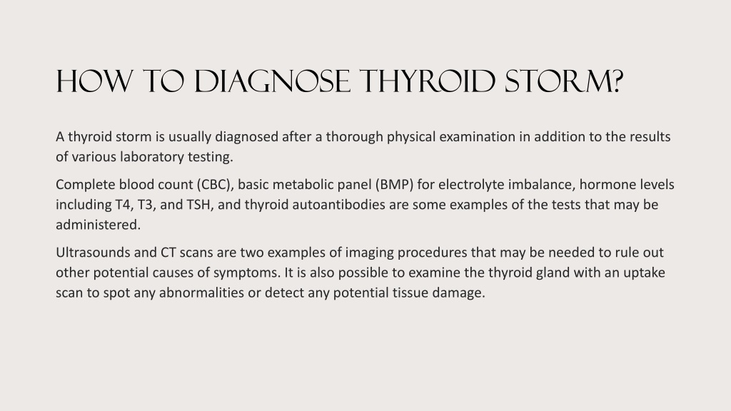
Thyroid function tests are essential in evaluating the overall thyroid gland function and detecting any underlying thyroid disorders that might be contributing to or mimicking thyroid stromal tumors. These tests measure the levels of thyroid hormones, such as thyroxine (T4) and triiodothyronine (T3), and thyroid-stimulating hormone (TSH).
- TSH: This hormone, produced by the pituitary gland, stimulates the thyroid gland to produce T4 and T3. Elevated TSH levels often indicate hypothyroidism, while low TSH levels suggest hyperthyroidism. In cases of thyroid stromal tumors, TSH levels may be altered depending on the tumor’s location and its impact on thyroid function.
- Free T4 and Free T3: These are the active forms of thyroid hormones that circulate in the bloodstream. Elevated free T4 and T3 levels indicate hyperthyroidism, while low levels suggest hypothyroidism. Analyzing these levels can help determine the extent of thyroid dysfunction associated with a thyroid stromal tumor.
Thyroid Ultrasound
Thyroid ultrasound is a non-invasive imaging technique that uses sound waves to create images of the thyroid gland. It provides detailed anatomical information about the thyroid gland, including its size, shape, and any nodules or masses present.
- Nodule Characterization: Ultrasound helps determine the characteristics of thyroid nodules, such as their size, shape, echogenicity (how the tissue reflects sound waves), and blood flow. These features can provide clues about the nature of the nodule, whether it is benign or malignant.
- Tumor Location and Size: Thyroid ultrasound helps identify the location and size of thyroid stromal tumors. This information is crucial for surgical planning and treatment.
- Fine-Needle Aspiration Biopsy (FNAB): Ultrasound-guided FNAB is a procedure that uses a thin needle to extract cells from a suspicious nodule. The extracted cells are then examined under a microscope by a pathologist to determine the type of tissue and whether it is cancerous.
Thyroid Scintigraphy
Thyroid scintigraphy, also known as a thyroid scan, is a nuclear medicine procedure that uses a radioactive tracer to visualize the thyroid gland. This technique can help determine the function of different parts of the thyroid gland and identify areas of abnormal activity.
- Hot Nodules: In thyroid scintigraphy, hot nodules appear as areas of increased uptake of the radioactive tracer, indicating increased thyroid hormone production. These nodules are usually benign, but further investigation is necessary to rule out thyroid cancer.
- Cold Nodules: Cold nodules, on the other hand, appear as areas of decreased tracer uptake, suggesting a lack of thyroid hormone production. Cold nodules are more likely to be malignant, but further investigation, such as FNAB, is needed for definitive diagnosis.
Computed Tomography (CT) Scan
CT scans are a type of imaging that uses X-rays to create detailed cross-sectional images of the body. CT scans can be helpful in evaluating the extent of thyroid stromal tumors and their relationship to surrounding structures.
- Tumor Staging: CT scans can help determine the stage of a thyroid stromal tumor, which refers to the tumor’s size, location, and spread to nearby lymph nodes or other organs.
- Surgical Planning: CT scans provide valuable information for surgical planning, allowing surgeons to identify the location of the tumor and plan the optimal surgical approach.
Magnetic Resonance Imaging (MRI)
MRI is a non-invasive imaging technique that uses magnetic fields and radio waves to create detailed images of the body. MRI is particularly useful in visualizing soft tissues, such as the thyroid gland.
- Tumor Characterization: MRI can help differentiate between different types of thyroid stromal tumors based on their signal intensity and contrast enhancement.
- Tumor Invasion: MRI can assess the extent of tumor invasion into surrounding tissues, which is important for determining the best treatment strategy.
Histopathology
Histopathology is the microscopic examination of tissue samples obtained through FNAB or surgery. This is the gold standard for diagnosing thyroid stromal tumors and determining their specific subtype.
- Tumor Classification: Histopathology allows pathologists to classify thyroid stromal tumors based on their cellular features, growth patterns, and presence of specific markers.
- Grading: Histopathological analysis helps determine the grade of a thyroid stromal tumor, which reflects its aggressiveness and potential for spread.
Molecular Testing
Molecular testing analyzes the genetic makeup of tumor cells to identify specific mutations or gene rearrangements that may be associated with thyroid stromal tumors.
- Prognostic Markers: Molecular testing can identify prognostic markers, which are genetic features that predict the likelihood of tumor recurrence or metastasis.
- Targeted Therapies: In some cases, molecular testing can identify specific mutations that make the tumor susceptible to targeted therapies, which are drugs that target specific molecular pathways involved in tumor growth.
Thyroid Function Tests (TFTs)
Thyroid function tests (TFTs) are a crucial component of the diagnostic workup for patients suspected of having thyroid stromal tumors. These tests assess the overall activity of the thyroid gland, providing valuable insights into its functional state and helping differentiate between various thyroid conditions.
Role of TFTs in Evaluating Thyroid Function
TFTs play a pivotal role in evaluating thyroid function in patients suspected of having thyroid stromal tumors. They provide a comprehensive assessment of the thyroid gland’s activity, aiding in the diagnosis and management of various thyroid disorders. TFTs measure the levels of thyroid hormones, including thyroxine (T4) and triiodothyronine (T3), as well as thyroid-stimulating hormone (TSH). These hormones work together to regulate metabolism, growth, and development.
Significance of Abnormal TFT Results
Abnormal TFT results can be highly significant in the context of thyroid stromal tumors.
- Elevated TSH levels, coupled with low T4 and T3 levels, may suggest hypothyroidism, a condition where the thyroid gland is not producing enough hormones.
- Conversely, suppressed TSH levels with high T4 and T3 levels may indicate hyperthyroidism, a condition characterized by excessive thyroid hormone production.
While thyroid stromal tumors are generally non-functional, meaning they do not directly impact thyroid hormone production, they can indirectly affect thyroid function by compressing or infiltrating the surrounding thyroid tissue.
Differentiating Thyroid Stromal Tumors from Other Thyroid Conditions
TFTs can help differentiate thyroid stromal tumors from other thyroid conditions. For example, a patient with a thyroid stromal tumor may present with normal TFT results, while a patient with Graves’ disease, an autoimmune disorder causing hyperthyroidism, would likely have elevated T4 and T3 levels and suppressed TSH levels.
TFTs can help rule out other thyroid conditions, such as thyroiditis, Hashimoto’s thyroiditis, and Graves’ disease, which can mimic the symptoms of thyroid stromal tumors.
TFTs, in conjunction with other diagnostic tests, such as ultrasound and fine-needle aspiration biopsy, are essential for accurately diagnosing thyroid stromal tumors and guiding appropriate treatment strategies.
Thyroid Ultrasound
Thyroid ultrasound is an essential imaging technique used in the diagnosis and management of thyroid stromal tumors. It provides valuable information about the size, shape, and location of the tumor, aiding in determining the extent of the tumor and guiding biopsy procedures for definitive diagnosis.
Visualization of Thyroid Stromal Tumors
Thyroid ultrasound utilizes high-frequency sound waves to create images of the thyroid gland. These images can clearly depict the presence of thyroid stromal tumors, which often appear as solid, hypoechoic (darker) masses within the thyroid tissue.
Assessing Tumor Size, Shape, and Location
Ultrasound allows for precise measurement of the tumor’s dimensions (length, width, and height). It also helps visualize the tumor’s shape, whether it is well-defined or irregular, and its location within the thyroid gland (e.g., right or left lobe, isthmus). This information is crucial for determining the stage of the tumor and planning appropriate treatment strategies.
Guidance for Biopsy Procedures
Thyroid ultrasound plays a critical role in guiding fine-needle aspiration biopsy (FNAB), a minimally invasive procedure used to obtain tissue samples from suspicious thyroid nodules. The ultrasound image allows the physician to accurately target the tumor and guide the needle for optimal sample collection.
FNAB is a highly accurate diagnostic tool for thyroid stromal tumors, providing a definitive diagnosis based on the microscopic examination of the collected cells.
Fine Needle Aspiration Biopsy (FNAB)
Fine needle aspiration biopsy (FNAB) is a minimally invasive procedure that involves using a thin needle to collect cells from a suspicious thyroid nodule for microscopic examination. It plays a crucial role in the diagnosis of thyroid stromal tumors, providing valuable information about the nature and characteristics of the tumor.FNAB is a safe and effective procedure that can be performed in an outpatient setting.
It typically involves the following steps:
Procedure of FNAB
- The patient lies down on an examination table, and the area of the thyroid nodule is cleaned and numbed with a local anesthetic.
- A thin needle attached to a syringe is inserted into the nodule, and a small amount of tissue is aspirated.
- The aspirated cells are then spread onto a glass slide and stained for microscopic examination by a pathologist.
Cytological Features of FNAB Specimens
The cytological features of FNAB specimens from thyroid stromal tumors can vary depending on the specific type of tumor. However, some common characteristics include:
- Spindle-shaped cells: These are elongated cells with a central nucleus and a small amount of cytoplasm. They are often arranged in a whorled or fascicular pattern.
- Myxoid stroma: This refers to the presence of a gelatinous, mucoid substance surrounding the tumor cells. It is often seen in myxoid tumors.
- Cellular atypia: This refers to abnormalities in the appearance of the tumor cells, such as enlarged nuclei, irregular shapes, and increased mitotic activity.
Determining Tumor Type and Grade
FNAB can help determine the type and grade of a thyroid stromal tumor by analyzing the cytological features of the aspirated cells. For example, a FNAB specimen showing spindle-shaped cells with a myxoid stroma and cellular atypia may be suggestive of a myxoid liposarcoma. The grade of the tumor, which indicates its aggressiveness, can be assessed by examining the degree of cellular atypia and the number of mitotic figures.
Other Diagnostic Tools: Are Thyroid Studies Necessary To Diagnose Thyroid Strom
While thyroid studies play a crucial role in evaluating thyroid stromal tumors, other imaging modalities can provide complementary information, contributing to a comprehensive diagnostic picture. These tools offer unique perspectives on the tumor’s size, location, and relationship with surrounding structures, aiding in treatment planning and follow-up monitoring.
Computed Tomography (CT) Scan
CT scans use X-rays and computer processing to create detailed cross-sectional images of the body. In the context of thyroid stromal tumors, CT scans can provide valuable information about the tumor’s size, shape, and extent of spread, particularly if it has extended beyond the thyroid gland. They can also help determine the involvement of nearby structures like the trachea, esophagus, and major blood vessels.
CT scans are particularly useful in visualizing bony structures and detecting calcifications within the tumor, which may be associated with certain subtypes of thyroid stromal tumors.
Magnetic Resonance Imaging (MRI)
MRI uses magnetic fields and radio waves to produce detailed images of soft tissues, making it a valuable tool for evaluating thyroid stromal tumors. MRI offers superior soft tissue contrast compared to CT, allowing for better visualization of the tumor’s internal structure and its relationship with surrounding tissues. It can also help distinguish between benign and malignant tumors based on the tumor’s signal intensity and the presence of certain features, such as capsule invasion or vascularity.
MRI is particularly useful in assessing the involvement of the larynx, which is a critical structure that may be affected by thyroid stromal tumors.
Positron Emission Tomography (PET) Scan
PET scans are used to detect metabolic activity in the body, making them particularly useful in identifying and characterizing tumors. By injecting a radioactive tracer that is taken up by metabolically active cells, PET scans can highlight areas of increased activity within the thyroid gland. While not routinely used in the initial diagnosis of thyroid stromal tumors, PET scans can be helpful in evaluating the extent of tumor spread, especially if there is suspicion of metastasis.
Role of Other Diagnostic Tools in the Overall Diagnostic Picture
The combination of thyroid studies, such as thyroid function tests, ultrasound, and FNAB, with other imaging modalities, such as CT, MRI, and PET, provides a comprehensive evaluation of thyroid stromal tumors. This multi-modal approach helps to accurately assess the tumor’s size, location, extent of spread, and potential involvement of surrounding structures. This information is crucial for determining the optimal treatment strategy and for monitoring the effectiveness of treatment.
Management of Thyroid Stromal Tumors
Managing thyroid stromal tumors involves a multidisciplinary approach tailored to the specific tumor type, stage, and patient’s overall health. The primary goals of treatment are to remove or control the tumor, prevent its spread, and maintain or improve the patient’s quality of life.
Surgical Intervention
Surgery is the mainstay of treatment for most thyroid stromal tumors. The extent of surgery depends on the tumor’s size, location, and the presence of lymph node involvement.
- Total thyroidectomy: This procedure involves the complete removal of the thyroid gland. It is often performed for larger tumors or those that have spread to nearby lymph nodes.
- Lobectomy: This procedure involves the removal of one lobe of the thyroid gland. It is typically considered for smaller tumors confined to one lobe.
- Lymph node dissection: This procedure involves the removal of lymph nodes in the neck to check for tumor spread and remove any affected nodes.
The success of surgery in managing thyroid stromal tumors depends on the tumor’s characteristics, the skill of the surgeon, and the patient’s overall health.
Radiation Therapy
Radiation therapy is often used in conjunction with surgery to treat thyroid stromal tumors, especially in cases where the tumor has spread to other parts of the body. Radiation therapy uses high-energy rays to damage and destroy cancer cells. It can be delivered externally using a machine or internally using radioactive implants.
- External beam radiation therapy: This type of radiation therapy delivers radiation to the tumor site from outside the body. It is commonly used to treat thyroid stromal tumors that have spread to nearby lymph nodes or other parts of the body.
- Radioactive iodine therapy: This type of radiation therapy uses a radioactive form of iodine that is taken orally. It is often used to treat thyroid cancer that has spread to other parts of the body.
Radiation therapy can be effective in controlling tumor growth and reducing symptoms. However, it can also cause side effects, such as fatigue, nausea, and hair loss.
Chemotherapy
Chemotherapy is a systemic treatment that uses drugs to kill cancer cells. It is less commonly used for thyroid stromal tumors than surgery or radiation therapy. Chemotherapy may be used to treat tumors that have spread to other parts of the body or to reduce the size of a tumor before surgery.
- Targeted therapy: These drugs target specific molecules involved in tumor growth and development.
- Immunotherapy: These drugs boost the body’s immune system to fight cancer cells.
Chemotherapy can cause side effects, such as nausea, vomiting, hair loss, and fatigue.
Prognosis and Long-Term Outcomes
The prognosis for patients with thyroid stromal tumors varies depending on the tumor type, stage, and the patient’s overall health.
- Early-stage tumors: Patients with early-stage tumors have a good prognosis. They often have a high chance of complete recovery with surgery alone.
- Advanced-stage tumors: Patients with advanced-stage tumors have a less favorable prognosis. They may require more aggressive treatment, such as radiation therapy or chemotherapy, and their chances of long-term survival may be lower.
The long-term outcomes for patients with thyroid stromal tumors are improving due to advances in diagnosis and treatment.
Understanding the importance of thyroid studies in diagnosing thyroid stromal tumors is essential for patients and healthcare providers. These studies provide valuable insights into the nature, extent, and potential treatment options for these tumors. While thyroid studies are not the only diagnostic tools employed, they play a crucial role in creating a comprehensive picture of the patient’s condition.
FAQ Insights
What are the symptoms of thyroid stromal tumors?
Symptoms can vary depending on the size and location of the tumor. Some common symptoms include a lump in the neck, difficulty swallowing, hoarseness, and pain in the neck.
What are the different types of thyroid stromal tumors?
There are several types of thyroid stromal tumors, each with unique characteristics. These include fibrosarcoma, malignant fibrous histiocytoma, and liposarcoma.
How are thyroid stromal tumors treated?
Treatment options for thyroid stromal tumors depend on the type, stage, and overall health of the patient. Treatment may include surgery, radiation therapy, or chemotherapy.
What is the prognosis for patients with thyroid stromal tumors?
The prognosis for patients with thyroid stromal tumors varies depending on the type and stage of the tumor. Early detection and treatment are crucial for improving outcomes.


