How does corneal stroma regeneration happen? This question delves into the fascinating world of the cornea, a transparent tissue that protects our eyes and allows us to see. The corneal stroma, its middle layer, plays a vital role in maintaining this clarity. But what happens when the stroma is injured? Can it heal itself, and if so, how?
This journey explores the intricate mechanisms behind corneal stromal regeneration, uncovering the remarkable abilities of the eye to repair itself.
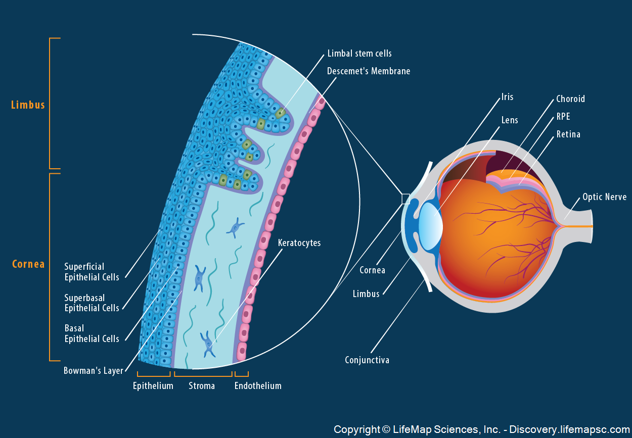
The corneal stroma, composed of a complex network of collagen fibers and specialized cells called keratocytes, acts as a foundation for corneal transparency. This unique structure allows light to pass through the cornea, enabling clear vision. However, the corneal stroma has limited regenerative capabilities compared to other tissues in the body, making its repair a complex process.
The Corneal Stroma

The corneal stroma is the central and thickest layer of the cornea, accounting for approximately 90% of its total thickness. It’s a remarkably organized and highly specialized tissue that plays a crucial role in maintaining the cornea’s transparency and refractive properties, allowing light to pass through the eye unimpeded.
Structure and Composition
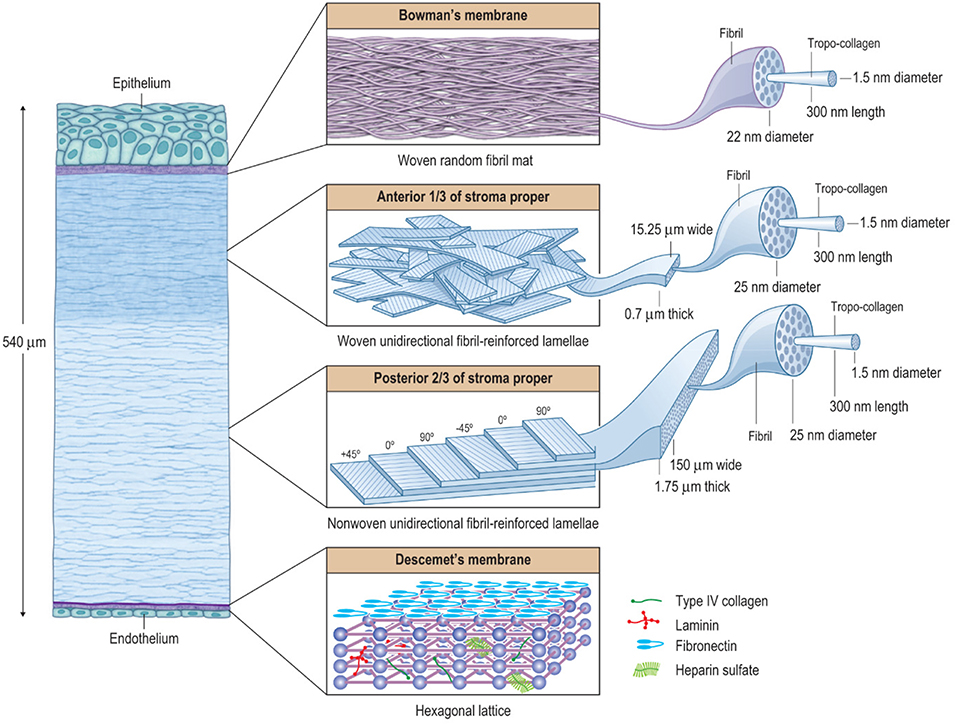
The corneal stroma is primarily composed of a dense, interwoven network of collagen fibrils embedded in a hydrated matrix of proteoglycans and water. This intricate structure is responsible for the cornea’s exceptional strength and resilience, while the high water content ensures its transparency.
Key Cell Types
The corneal stroma is populated by various cell types, each playing a distinct role in maintaining its integrity and function.
- Keratocytes: These are the primary resident cells of the stroma, responsible for synthesizing and maintaining the collagen fibrils and other extracellular matrix components. They are responsible for the structural integrity of the stroma.
- Fibroblasts: These cells are involved in wound healing and tissue repair. They can differentiate into myofibroblasts, contributing to the contraction of the wound site.
- Immune cells: The stroma also contains a small population of immune cells, such as macrophages and mast cells, which play a role in defense against infection and inflammation.
Organization of Collagen Fibrils
The collagen fibrils in the stroma are arranged in a highly organized, parallel fashion, forming lamellae that are stacked on top of each other. This unique organization is critical for the cornea’s transparency. The tightly packed collagen fibrils with their uniform diameter and spacing ensure that light passes through the stroma with minimal scattering. The regular arrangement of the fibrils prevents light from being deflected in multiple directions, maintaining the cornea’s transparency.
The corneal stroma’s unique organization, with its densely packed and regularly arranged collagen fibrils, is analogous to a carefully constructed optical fiber, effectively channeling light through the cornea.
The Limits of Corneal Regeneration
While the corneal stroma exhibits remarkable regenerative capabilities, it is not without limitations. Unlike some other tissues in the body, the stroma’s ability to repair itself is not limitless, and factors such as the nature of the injury, the age of the individual, and the presence of underlying health conditions can significantly influence the outcome of regeneration.
The Role of Quiescent Stromal Cells
The corneal stroma’s limited regenerative capacity is largely attributed to the presence of a quiescent stromal cell population. These cells, known as keratocytes, are responsible for maintaining the structural integrity of the stroma. While they have the potential to differentiate into other cell types, they normally exist in a resting state, only becoming active in response to injury or other stimuli.
This quiescent nature of keratocytes limits the rate and extent of stromal regeneration.
The Lack of Significant Vascularization
Another factor that contributes to the limited regenerative capacity of the corneal stroma is the lack of significant vascularization. Unlike many other tissues, the corneal stroma is avascular, meaning it does not have a direct blood supply. This absence of blood vessels restricts the delivery of nutrients, growth factors, and other essential molecules necessary for tissue repair and regeneration.
Consequently, the stroma relies on diffusion from surrounding tissues for its metabolic needs, which can limit the rate of cell proliferation and wound healing.
Comparison with Other Tissues
The regenerative potential of the corneal stroma contrasts with that of other tissues in the body, such as skin and bone. Skin, being a highly vascularized tissue, possesses a significant regenerative capacity. Its epithelial layer constantly renews itself, and fibroblasts within the dermis readily proliferate to repair wounds. Bone, while possessing a slower regeneration rate, is capable of significant repair and remodeling due to the presence of osteoblasts and osteoclasts.
The Role of Keratocytes in Regeneration

Keratocytes, the primary resident cells of the corneal stroma, play a crucial role in maintaining corneal transparency and actively participate in wound healing and regeneration. Their ability to respond to injury and initiate the repair process makes them central to corneal homeostasis.
Types of Keratocytes and Their Roles
Keratocytes exhibit diverse morphologies and functional characteristics, influencing their roles in corneal regeneration.
- Myofibroblasts: These keratocytes, characterized by their elongated shape and the presence of contractile proteins like α-smooth muscle actin (α-SMA), contribute to wound closure by contracting the stroma. They also play a role in collagen synthesis, contributing to the formation of a scar tissue.
- Fibroblasts: These keratocytes are responsible for synthesizing and depositing extracellular matrix (ECM) components, such as collagen, which forms the structural framework of the cornea. Their role is essential for maintaining corneal transparency and restoring the damaged stroma.
- Quiescent Keratocytes: These cells are the predominant type in the healthy cornea, characterized by their flattened morphology and low metabolic activity. They maintain the corneal stroma’s integrity and transparency. In response to injury, they become activated and transform into myofibroblasts or fibroblasts.
Activation of Keratocytes in Response to Injury, How does corneal stroma regeneration happen
Upon injury, keratocytes undergo a complex activation process, involving signaling pathways and molecular events.
- Growth Factors: Injury triggers the release of growth factors, such as platelet-derived growth factor (PDGF) and transforming growth factor-β (TGF-β), which stimulate keratocyte proliferation and differentiation.
- Cytokines: These signaling molecules, released by various cells, activate keratocytes and induce their transformation into myofibroblasts or fibroblasts.
- Extracellular Matrix Remodeling: Injury disrupts the corneal stroma’s ECM, triggering the activation of keratocytes and initiating the repair process.
Differentiation Potential of Keratocytes
Keratocytes exhibit a remarkable plasticity, enabling them to differentiate into other cell types, contributing to the regeneration process.
- Fibroblasts: Keratocytes can differentiate into fibroblasts, which synthesize and deposit ECM components, essential for restoring the corneal stroma’s structure and transparency.
- Myofibroblasts: Keratocytes can transform into myofibroblasts, contributing to wound closure by contracting the stroma. However, this process can also lead to scar formation, compromising corneal transparency.
The Influence of the Corneal Epithelium

The corneal epithelium, the outermost layer of the cornea, plays a vital role in maintaining corneal integrity and promoting stromal regeneration. This transparent, multilayered structure acts as a barrier against external insults, while also actively participating in the healing process after injury.
Epithelial Cell Contributions to Stromal Regeneration
The corneal epithelium is not merely a passive barrier; it actively contributes to stromal regeneration by releasing a variety of growth factors and cytokines that stimulate keratocyte activity.
- Epithelial Growth Factor (EGF): This potent mitogen promotes keratocyte proliferation and differentiation, contributing to the formation of new stromal tissue.
- Transforming Growth Factor-beta (TGF-β): TGF-β plays a crucial role in wound healing, stimulating the production of extracellular matrix components, including collagen, which are essential for restoring corneal structure.
- Fibroblast Growth Factor (FGF): FGFs are a family of growth factors that promote angiogenesis (new blood vessel formation), which is vital for delivering nutrients and oxygen to the regenerating stroma.
These growth factors and cytokines create a signaling cascade that orchestrates the complex process of stromal regeneration.
Impact of Epithelial Defects on Stromal Regeneration
Epithelial defects or abnormalities can significantly hinder stromal regeneration.
- Delayed Healing: When the epithelial barrier is compromised, it delays the healing process, as the underlying stroma is exposed to external insults and the release of growth factors is disrupted.
- Scarring: Chronic epithelial defects or persistent inflammation can lead to excessive collagen deposition, resulting in scar formation and impaired vision.
- Infection: A compromised epithelial barrier increases the risk of infection, which can further complicate the healing process and damage the corneal stroma.
Therefore, maintaining the integrity of the corneal epithelium is essential for successful stromal regeneration.
Emerging Technologies for Corneal Regeneration
The quest to restore corneal function in cases of severe damage or disease has led to the development of several promising technologies that aim to enhance stromal regeneration. These technologies leverage the body’s inherent healing capabilities by providing scaffolds, stimulating cell growth, or directly modifying the genetic blueprint of corneal cells.
Comparison of Technologies for Enhancing Corneal Stromal Regeneration
Different approaches have been explored to stimulate corneal stromal regeneration, each with its own advantages and limitations. The following table provides a comparative analysis of these techniques:
| Approach | Description | Advantages | Disadvantages |
|---|---|---|---|
| Stem Cell Transplantation | Involves transplanting corneal limbal stem cells, which can differentiate into various corneal cell types, including keratocytes, to regenerate the stroma. |
|
|
| Biomaterial Scaffolds | Utilizes synthetic or natural materials to create a three-dimensional framework that supports stromal cell growth and organization. |
|
|
| Gene Therapy | Delivers therapeutic genes to corneal cells to enhance their regenerative potential, such as by stimulating keratocyte proliferation or promoting collagen production. |
|
|
| Growth Factor Delivery | Involves delivering growth factors, such as fibroblast growth factor (FGF) or platelet-derived growth factor (PDGF), to the corneal stroma to stimulate keratocyte proliferation and collagen synthesis. |
|
|
Future Directions for Research in Corneal Stromal Regeneration
Ongoing research in corneal stromal regeneration aims to address the limitations of current approaches and develop novel strategies for restoring corneal function.
- Development of Novel Biomaterials with Enhanced Biocompatibility and Regenerative Properties: Researchers are exploring the use of biomaterials that closely mimic the native corneal microenvironment, promoting cell adhesion, proliferation, and differentiation. Examples include biomaterials derived from natural sources like collagen or hyaluronic acid, as well as synthetic polymers with tailored properties.
- Engineering Strategies for Promoting Angiogenesis and Vascularization of the Corneal Stroma: The corneal stroma is typically avascular, which can hinder regeneration. Research is focused on developing strategies to induce angiogenesis in the stroma, improving nutrient and oxygen supply to the regenerating tissue. This can involve incorporating pro-angiogenic factors into biomaterials or using gene therapy to enhance vascularization.
- Exploring the Potential of Gene Editing Technologies to Enhance Stromal Regeneration: Gene editing technologies, such as CRISPR-Cas9, offer the possibility of precisely modifying the genetic code of corneal cells to enhance their regenerative potential. This could involve correcting genetic defects that contribute to corneal disease or introducing genes that promote stromal cell proliferation and collagen synthesis.
Understanding the intricacies of corneal stroma regeneration is crucial for developing innovative treatments for corneal injuries and diseases. Researchers are actively exploring the potential of stem cells, biomaterials, gene therapy, and growth factors to enhance this process. By harnessing the power of these emerging technologies, we can strive to restore sight and improve the quality of life for individuals facing corneal damage.
The future of corneal regeneration holds immense promise, offering hope for a world where vision is preserved and restored.
User Queries: How Does Corneal Stroma Regeneration Happen
What are the most common causes of corneal stroma damage?
Corneal stroma damage can occur due to various factors, including trauma, infections, and degenerative diseases. Common causes include corneal abrasions, ulcers, keratoconus, and Fuchs’ endothelial dystrophy.
Can corneal stromal regeneration be improved?
Yes, ongoing research is exploring various approaches to enhance corneal stromal regeneration. These include stem cell transplantation, biomaterial scaffolds, gene therapy, and growth factor delivery. These advancements hold promise for improving treatment outcomes for corneal injuries and diseases.


