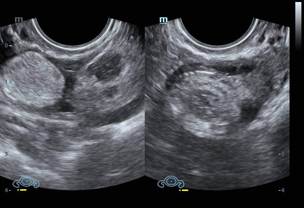
What is hyperechoic stroma? Imagine looking at a map with different colors representing different terrains. Ultrasound imaging works similarly, using sound waves to create images of the body’s internal structures. In this “map,” hyperechoic stroma appears as bright, white areas, indicating dense tissue. This density can be a sign of normal tissue or a potential indicator of various conditions, making understanding hyperechoic stroma crucial for accurate diagnosis and treatment.
Stroma, essentially the supporting framework of tissues and organs, plays a vital role in their structure and function. When stroma becomes hyperechoic, it signifies a change in its composition, which can be due to various factors, including inflammation, fibrosis, or even benign growths. Understanding the underlying causes and clinical implications of hyperechoic stroma is essential for medical professionals to provide appropriate care and guidance.
Introduction to Hyperechoic Stroma

Hyperechoic stroma refers to a tissue structure that appears brighter than surrounding tissues on ultrasound imaging. It’s like seeing a white patch against a gray background. This brightness indicates that sound waves are reflecting back strongly from that area, suggesting a denser or more solid structure. The significance of hyperechoic stroma in medical imaging lies in its ability to help identify various conditions.
This increased echogenicity can be a sign of normal tissue, like fibrous tissue or dense muscle, but it can also be a sign of disease, like fibrosis, calcification, or tumors.
Role of Stroma in Tissues and Organs, What is hyperechoic stroma
Stroma is the supporting framework of tissues and organs. It provides structure, support, and a pathway for blood vessels and nerves. In various organs, stroma can consist of different types of tissue, including connective tissue, fibrous tissue, and adipose tissue. For instance, in the breast, the stroma consists of fibrous tissue that supports the milk ducts and lobules. In the kidney, the stroma comprises connective tissue that surrounds the nephrons, the functional units of the kidney.
The composition and appearance of the stroma can vary depending on the organ and its specific function.
Characteristics of Hyperechoic Stroma: What Is Hyperechoic Stroma

Hyperechoic stroma is a common finding on ultrasound imaging, and its appearance can provide valuable information about the underlying tissue. Understanding the characteristics of hyperechoic stroma is essential for accurate interpretation of ultrasound images.
Appearance of Hyperechoic Stroma on Ultrasound Images
Hyperechoic stroma appears as bright, white areas on ultrasound images. This is because the sound waves are reflected back to the transducer more readily from dense tissues, such as fibrous tissue, collagen, and calcium deposits. In contrast, hypoechoic structures, such as fluid-filled cysts, appear darker because sound waves pass through them more easily.
Comparison of Hyperechoic Stroma with Other Echogenic Structures
It is important to differentiate hyperechoic stroma from other echogenic structures that may be present in the same area. For example, hyperechoic stroma may be mistaken for calcifications, which are also bright on ultrasound images. However, calcifications typically have a more sharply defined, angular appearance, whereas hyperechoic stroma may have a more diffuse, irregular appearance.
Factors Influencing the Echogenicity of Stroma
Several factors can influence the echogenicity of stroma, including:
- Tissue Density: Denser tissues, such as fibrous tissue and collagen, are more echogenic than less dense tissues, such as fat.
- Tissue Composition: The presence of calcium deposits, which are highly echogenic, can increase the echogenicity of stroma.
- Ultrasound Frequency: Higher frequency ultrasound probes produce images with better resolution but may also increase the echogenicity of some tissues.
- Ultrasound Technique: The angle of the ultrasound beam and the amount of pressure applied to the transducer can also influence the echogenicity of stroma.
Causes of Hyperechoic Stroma
Hyperechoic stroma is a common finding on ultrasound imaging, and its presence can be indicative of various underlying conditions. Understanding the causes of hyperechoic stroma is crucial for accurate diagnosis and appropriate management. This section will explore the common causes of hyperechoic stroma in different organs and tissues, the underlying mechanisms that contribute to increased echogenicity, and specific conditions associated with this finding.
Common Causes of Hyperechoic Stroma
Hyperechoic stroma can be caused by a variety of factors, including:
- Increased collagen deposition: Collagen is a fibrous protein that provides structural support to tissues. Increased collagen deposition can lead to increased echogenicity, as sound waves are reflected more readily by dense collagen fibers. This is commonly seen in conditions such as fibrosis, scarring, and chronic inflammation.
- Increased cellular density: A higher concentration of cells in the stroma can also contribute to increased echogenicity. This is because sound waves encounter more resistance as they travel through a denser tissue. This is often observed in tumors, inflammatory conditions, and some benign growths.
- Deposition of calcium: Calcium deposits, also known as calcifications, can be seen in the stroma of various tissues. These deposits are highly reflective of sound waves, resulting in a hyperechoic appearance. Calcifications can be associated with a variety of conditions, including chronic inflammation, trauma, and certain types of tumors.
- Fat infiltration: Fat is another highly echogenic tissue. In some cases, fat infiltration into the stroma can lead to increased echogenicity. This is commonly seen in conditions such as fatty liver disease and breast fibroadenomas.
- Fluid accumulation: Although fluid is typically anechoic (black) on ultrasound, the presence of small, scattered fluid collections within the stroma can appear hyperechoic due to the scattering of sound waves. This is often seen in inflammatory conditions and certain types of cysts.
Examples of Conditions Associated with Hyperechoic Stroma
Hyperechoic stroma is a common finding in a wide range of conditions. Here are some examples:
- Fibroids in the uterus: Fibroids are benign tumors that can occur in the uterus. They are often characterized by increased collagen deposition, leading to a hyperechoic appearance on ultrasound.
- Breast fibroadenomas: Fibroadenomas are benign tumors that can occur in the breast. They are often characterized by increased cellular density and sometimes fat infiltration, leading to a hyperechoic appearance on ultrasound.
- Chronic pancreatitis: Chronic pancreatitis is an inflammatory condition that can lead to fibrosis and calcifications in the pancreas, resulting in hyperechoic stroma on ultrasound.
- Prostate cancer: Prostate cancer can be associated with increased cellular density and calcifications in the prostate gland, leading to hyperechoic stroma on ultrasound.
- Kidney stones: Kidney stones are calcifications that can occur in the kidneys. They are highly echogenic and can be easily visualized on ultrasound.
Clinical Implications of Hyperechoic Stroma

The presence of hyperechoic stroma on ultrasound imaging can have significant clinical implications, depending on the organ or tissue being examined. It can serve as a diagnostic marker, influencing treatment decisions and impacting the overall prognosis.
Hyperechoic Stroma as a Diagnostic Marker
The presence of hyperechoic stroma can be indicative of various conditions, making it a valuable diagnostic marker. It can help differentiate between benign and malignant lesions, guide biopsy procedures, and monitor disease progression.
- Breast Cancer: In breast imaging, hyperechoic stroma can be associated with invasive ductal carcinoma, particularly in the presence of microcalcifications. This finding can prompt further investigation, such as a biopsy, to confirm the diagnosis.
- Prostate Cancer: Hyperechoic stroma in the prostate can be a sign of benign prostatic hyperplasia (BPH), a common condition characterized by an enlarged prostate. However, it can also be associated with prostate cancer, particularly in men with a family history of the disease.
- Ovarian Cysts: In ovarian imaging, hyperechoic stroma within a cyst can indicate a solid component, suggesting a potentially malignant tumor. This finding requires further evaluation to determine the nature of the cyst and the appropriate course of action.
Impact on Treatment Decisions
The presence of hyperechoic stroma can significantly influence treatment decisions. For example, in breast cancer, the presence of hyperechoic stroma can indicate a more aggressive tumor, potentially requiring more aggressive treatment modalities. In prostate cancer, hyperechoic stroma can help determine the extent of the disease and guide treatment options, such as surgery or radiation therapy.
Monitoring Disease Progression
Hyperechoic stroma can also be used to monitor disease progression. In cases of fibroids, for example, the presence of hyperechoic stroma can indicate growth or changes in the fibroid’s structure. This information can help guide treatment decisions and monitor the effectiveness of therapy.
In the intricate world of medical imaging, hyperechoic stroma serves as a valuable clue, often guiding clinicians towards a deeper understanding of the underlying health conditions. By recognizing its appearance, understanding its potential causes, and appreciating its clinical significance, we gain a clearer picture of the body’s inner workings. Remember, hyperechoic stroma is a signpost, prompting further investigation and ultimately leading to better patient care.
Query Resolution
What are some common causes of hyperechoic stroma in the breast?
Hyperechoic stroma in the breast can be caused by various factors, including fibrocystic changes, benign tumors, and even early stages of breast cancer. Further investigation with additional imaging or biopsy is often necessary for a definitive diagnosis.
Can hyperechoic stroma be seen in other organs besides the breast?
Yes, hyperechoic stroma can be observed in various organs, including the prostate, ovaries, kidneys, and liver. The specific causes and clinical implications may vary depending on the organ involved.
Is hyperechoic stroma always a sign of something serious?
Not necessarily. Hyperechoic stroma can sometimes be a normal finding, especially in younger individuals. However, it is important to consider the patient’s history, other clinical findings, and additional imaging to determine the significance of hyperechoic stroma.


