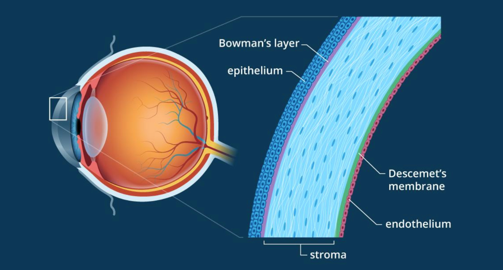
What is the corneal stroma? It’s the transparent, middle layer of the cornea, the front part of the eye. This seemingly simple structure plays a crucial role in maintaining the eye’s shape and allowing light to pass through, ultimately enabling us to see. The corneal stroma is a marvel of biological engineering, with its intricate arrangement of collagen fibrils and keratocytes ensuring both strength and transparency.
This delicate balance is essential for clear vision, and any disruption to the stromal structure can lead to visual impairment.
Imagine the cornea as a window to the world. The corneal stroma acts as the glass, providing the structural support and clarity necessary for light to pass through and reach the retina. This intricate layer is made up of a dense network of collagen fibers, arranged in a precise and organized fashion. These fibers are interwoven with specialized cells called keratocytes, which play a crucial role in maintaining the stroma’s integrity and ensuring its transparency.
The corneal stroma’s unique composition and structure make it a fascinating area of study, with implications for understanding vision, treating eye diseases, and developing new regenerative therapies.
The Corneal Stroma
The corneal stroma is the central and thickest layer of the cornea, playing a crucial role in maintaining the eye’s shape and transparency. It is a complex and highly organized structure, responsible for providing the cornea with its structural integrity and allowing for the passage of light.
The Corneal Stroma: Composition and Structure
The corneal stroma is primarily composed of collagen fibrils, embedded in a matrix of proteoglycans and water. These collagen fibrils are arranged in a highly organized, parallel fashion, forming lamellae. Each lamella is made up of bundles of collagen fibrils, all aligned in the same direction, but with adjacent lamellae oriented at slightly different angles. This unique arrangement gives the stroma its remarkable strength and flexibility.
- Collagen fibrils: These fibrils are made up of the protein collagen, specifically type I collagen. The collagen molecules self-assemble into fibrils, which then form larger bundles. The arrangement of these fibrils is crucial for the cornea’s transparency and structural integrity.
- Keratocytes: These cells are responsible for synthesizing and maintaining the collagen matrix of the stroma. They reside between the lamellae, playing a vital role in the cornea’s repair and regeneration processes.
- Proteoglycans: These molecules are responsible for attracting and retaining water within the stroma. They help to maintain the hydration of the cornea, which is essential for its transparency and flexibility.
The Importance of the Stromal’s Organized Structure
The highly organized structure of the corneal stroma is essential for its function in light transmission. The parallel arrangement of collagen fibrils minimizes light scattering, allowing light to pass through the cornea with minimal distortion. This is crucial for clear vision.
The organized structure of the corneal stroma, with its precisely aligned collagen fibrils, acts like a natural lens, ensuring that light rays are refracted and focused correctly onto the retina.
Collagen Fibrils
The corneal stroma, the middle layer of the cornea, is primarily composed of collagen fibrils, which are long, thin fibers made up of the protein collagen. These fibrils are arranged in a highly organized and precise manner, contributing significantly to the cornea’s remarkable transparency and structural integrity.
Unique Properties of Collagen Fibrils in the Corneal Stroma
The arrangement and spacing of collagen fibrils in the corneal stroma are crucial for its function. The fibrils are aligned in parallel lamellae, thin sheets stacked on top of each other, with a precise spacing between them. This arrangement allows for the passage of light with minimal scattering, ensuring the cornea’s transparency. The precise spacing between the fibrils also contributes to the cornea’s tensile strength and resistance to deformation, enabling it to withstand the pressure of the eye.
Comparison of Collagen Fibrils in Different Tissues
Collagen fibrils are found in various tissues throughout the body, each with a unique structure and function. In the corneal stroma, the collagen fibrils are exceptionally thin and tightly packed, with a uniform diameter and spacing. This arrangement is distinct from collagen fibrils in other tissues, such as tendons and ligaments, where the fibrils are thicker and more loosely packed.
The specific arrangement of collagen fibrils in the cornea is essential for its unique optical and mechanical properties.
Role of Proteoglycans and Water in Maintaining the Integrity of the Corneal Stroma
Proteoglycans, large molecules composed of protein and sugar chains, are embedded within the corneal stroma. These molecules, along with water, play a crucial role in maintaining the integrity and hydration of the corneal stroma. The proteoglycans form a hydrated gel-like matrix that surrounds the collagen fibrils, contributing to the cornea’s resilience and flexibility. The water content of the stroma is essential for maintaining its transparency and refractive index, allowing light to pass through without scattering.
Keratocytes
The corneal stroma, the transparent and robust middle layer of the cornea, is home to a specialized population of cells known as keratocytes. These cells play a crucial role in maintaining the structural integrity and transparency of the cornea. They are responsible for synthesizing and organizing the collagen fibrils that form the stroma’s framework, ensuring its resilience and clarity.
Morphology and Function of Keratocytes, What is the corneal stroma
Keratocytes are stellate-shaped cells with long, thin processes that extend throughout the stroma. Their morphology reflects their function, as these processes enable them to interact with the collagen fibrils and maintain the stroma’s organized structure. Keratocytes are highly specialized cells that primarily synthesize and maintain the collagenous matrix of the stroma. Their cytoplasm contains abundant rough endoplasmic reticulum and Golgi apparatus, organelles crucial for protein synthesis and secretion.Keratocytes synthesize a variety of extracellular matrix (ECM) components, including collagen types I, III, V, and VI, as well as proteoglycans and glycoproteins.
These components contribute to the stroma’s tensile strength, transparency, and hydration. The collagen fibrils produced by keratocytes are precisely arranged in a parallel fashion, forming lamellae that are stacked upon each other. This precise organization allows for the efficient transmission of light through the cornea, maintaining its transparency.Keratocytes also play a crucial role in stromal homeostasis. They continuously monitor and adjust the ECM composition and structure, ensuring the stroma’s integrity and functionality.
They can degrade and remodel the ECM, removing damaged or aged components, and replacing them with newly synthesized molecules. This constant turnover ensures the stroma’s structural integrity and transparency over time.
The Stroma’s Role in Vision: What Is The Corneal Stroma

The corneal stroma, the thickest layer of the cornea, plays a crucial role in vision. Its unique structure and composition contribute significantly to the eye’s ability to focus light and maintain visual clarity.
The Stroma’s Refractive Index
The stroma’s refractive index, a measure of how much light bends as it passes through the cornea, is critical for focusing light onto the retina. The tightly packed collagen fibrils and the aqueous humor within the stroma create a refractive index slightly higher than that of air. This difference in refractive index allows the cornea to bend light rays, bringing them into focus on the retina.
The Impact of Stromal Abnormalities on Vision
Any disruption to the stroma’s structure can impair vision.
- Corneal Edema: When the stroma becomes swollen, it can cause the cornea to bulge outward, distorting the shape of the cornea and leading to blurry vision. This can occur due to various factors, including inflammation, trauma, or certain medical conditions.
- Corneal Scarring: Scars in the stroma can also disrupt the cornea’s shape and clarity, causing blurred or distorted vision. Scars can result from injuries, infections, or surgical procedures.
Maintaining Stromal Transparency
The remarkable transparency of the corneal stroma is essential for clear vision.
- Keratocytes: These specialized cells within the stroma maintain the stroma’s structure and transparency. Keratocytes produce and maintain the collagen fibrils, ensuring their proper arrangement and spacing. They also play a role in wound healing and scar formation.
- Collagen Fibrils: The parallel arrangement of collagen fibrils in the stroma allows light to pass through with minimal scattering. The precise spacing and organization of these fibrils contribute to the cornea’s transparency.
Clinical Relevance of the Corneal Stroma

The corneal stroma, as the primary structural component of the cornea, plays a crucial role in maintaining the eye’s transparency and refractive power. However, its intricate structure and delicate balance are susceptible to various diseases and disorders, impacting vision and requiring clinical intervention. Understanding these conditions and their treatments is vital for effective ophthalmological care.
Corneal Stromal Diseases and Disorders
Corneal stromal diseases encompass a range of conditions affecting the stroma’s structure and function, leading to impaired vision. These conditions can be broadly categorized into two main groups:
- Keratoconus: A progressive corneal disorder characterized by thinning and protrusion of the cornea, leading to irregular astigmatism and distorted vision. This condition often results from a genetic predisposition, with weakened collagen fibrils in the stroma being a key factor.
- Corneal Dystrophies: A group of inherited disorders that affect the corneal stroma’s structure and composition. These dystrophies can manifest as various symptoms, including corneal opacities, scarring, and impaired vision. Different types of corneal dystrophies are associated with specific genetic mutations, leading to abnormal collagen production or deposition.
Surgical Procedures for Stromal Abnormalities
Surgical interventions are often necessary to address stromal abnormalities and restore visual function. Two primary surgical approaches are commonly employed:
- Corneal Transplantation: A surgical procedure involving the replacement of a damaged cornea with a healthy donor cornea. This procedure is often used for severe corneal diseases, such as keratoconus, corneal dystrophies, and corneal scarring.
- Refractive Surgery: A range of surgical procedures aimed at correcting refractive errors, such as myopia, hyperopia, and astigmatism. These procedures often involve reshaping the corneal stroma using lasers or other techniques to improve visual acuity.
Stem Cell Therapy and Gene Therapy
Emerging technologies like stem cell therapy and gene therapy hold significant promise for treating corneal stromal diseases and enhancing corneal regeneration.
- Stem Cell Therapy: Utilizes stem cells, which have the potential to differentiate into various cell types, including corneal stromal cells. Stem cell transplantation can potentially regenerate damaged corneal tissue and restore its function.
- Gene Therapy: Aims to correct genetic defects underlying certain corneal dystrophies by delivering therapeutic genes to the corneal cells. This approach has the potential to address the root cause of the disease and prevent further progression.
The corneal stroma is a testament to the intricate design of the human eye. Its remarkable structure and function allow us to see the world around us with clarity. Understanding the stroma’s complexities is essential for addressing corneal diseases and developing innovative therapies to restore vision. From the precise arrangement of collagen fibers to the vital role of keratocytes, the corneal stroma is a fascinating example of how nature combines strength, transparency, and cellular activity to achieve a vital function.
Popular Questions
What is the difference between the corneal stroma and the corneal endothelium?
The corneal stroma is the middle layer of the cornea, while the corneal endothelium is the innermost layer. The endothelium is a single layer of cells that regulates the flow of fluid into and out of the stroma, maintaining its hydration and transparency.
What are the most common corneal stromal diseases?
Common corneal stromal diseases include keratoconus, a condition that causes the cornea to thin and bulge, and corneal dystrophies, which are genetic disorders that affect the structure and function of the stroma.
How does corneal transplantation work?
Corneal transplantation involves replacing the damaged or diseased cornea with a healthy donor cornea. The procedure can restore vision in patients with severe corneal disease.


