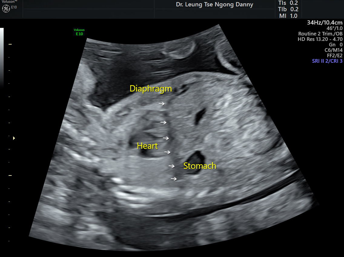
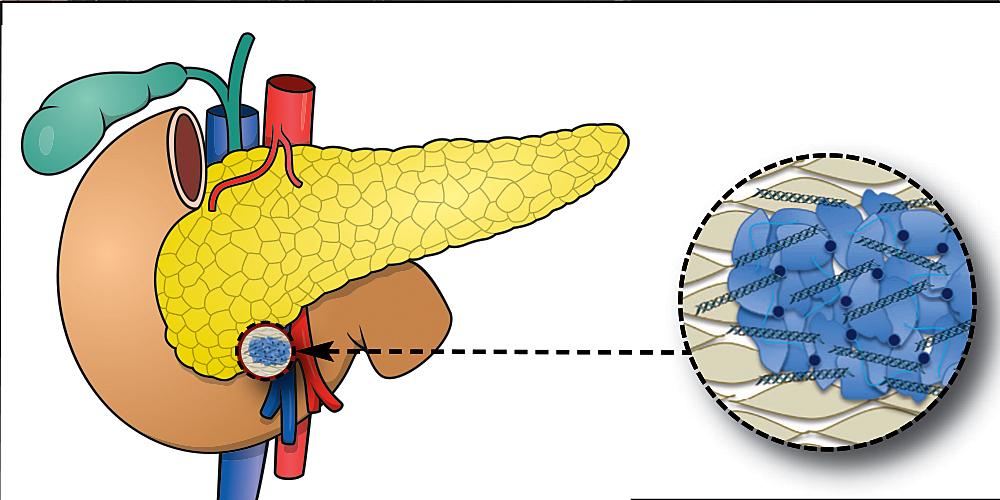
What is the meaning of echogenic stroma? This seemingly simple question delves into a complex world of medical imaging, where echoes on ultrasound scans reveal intricate details about our bodies. Echogenic stroma, characterized by bright, white areas on ultrasound images, often signifies dense tissue. This density can arise from various causes, ranging from normal tissue variations to underlying medical conditions.
Understanding the meaning of echogenic stroma is crucial for healthcare professionals to accurately diagnose and manage patient care.
Ultrasound imaging, a non-invasive technique that utilizes sound waves to create images of internal organs, plays a pivotal role in revealing echogenic stroma. By understanding the principles of ultrasound and the relationship between echogenicity and tissue density, we can gain valuable insights into the significance of echogenic stroma in different organs and medical conditions.
Introduction to Echogenic Stroma
Echogenic stroma is a term used in medical imaging, particularly in ultrasound scans, to describe an area within an organ or tissue that appears brighter than the surrounding tissue. This brightness is due to the reflection of sound waves, which are used to create the ultrasound image. In simpler terms, echogenic stroma is a region that shows up as white or very light gray on an ultrasound.
Examples of Medical Conditions, What is the meaning of echogenic stroma
Echogenic stroma can be observed in various medical conditions, often indicating changes in the tissue structure. Here are some examples:
- Fibroids: These are noncancerous growths in the uterus. They can appear echogenic on ultrasound due to the presence of dense fibrous tissue.
- Endometriosis: This condition involves the growth of endometrial tissue outside the uterus. Echogenic stroma can be seen in endometriosis lesions, particularly in the ovaries.
- Ovarian cysts: Some ovarian cysts, such as dermoid cysts, can have echogenic components due to the presence of hair, teeth, or other tissues within the cyst.
- Breast cancer: While not always the case, some breast cancers can appear echogenic on ultrasound, especially if they are dense or contain calcium deposits.
- Chronic pancreatitis: This condition involves inflammation of the pancreas. Echogenic stroma can be seen in the pancreas due to scarring and fibrosis.
It’s important to note that echogenic stroma alone is not always a cause for concern. However, it can be a sign of underlying medical conditions. If you have any concerns, it’s crucial to consult with a doctor for further evaluation and diagnosis.
Echogenicity and Ultrasound Imaging

Ultrasound imaging, also known as sonography, is a non-invasive medical imaging technique that uses high-frequency sound waves to create images of internal organs and structures. This technology is widely used for various medical purposes, including diagnosing and monitoring various conditions, guiding procedures, and providing real-time visualization of internal organs.
Echogenicity Measurement and Representation
Echogenicity is a crucial concept in ultrasound imaging, referring to the ability of tissues and structures to reflect sound waves. Different tissues have varying densities and compositions, leading to different levels of echogenicity. Ultrasound images are generated based on the strength of the reflected sound waves.
- High echogenicity: Tissues with high echogenicity reflect sound waves strongly, appearing bright or white on the ultrasound image. These tissues are typically dense, such as bones, calcifications, or fibrous tissues.
- Low echogenicity: Tissues with low echogenicity reflect sound waves weakly, appearing dark or black on the ultrasound image. These tissues are typically less dense, such as fluids, cysts, or fat.
- Isoechogenicity: Tissues with similar echogenicity to the surrounding tissues appear the same shade of gray on the ultrasound image.
Relationship between Echogenicity and Tissue Density
Echogenicity is directly related to the density of tissues. Dense tissues, such as bones, reflect sound waves more effectively, resulting in high echogenicity. In contrast, less dense tissues, such as fluids, reflect sound waves poorly, leading to low echogenicity.
The relationship between echogenicity and tissue density is a fundamental principle in ultrasound imaging, allowing healthcare professionals to distinguish different tissues and structures based on their appearance on ultrasound images.
Causes of Echogenic Stroma

Echogenic stroma, as we have learned, is a common finding in ultrasound imaging. It signifies increased echogenicity in the connective tissue supporting various organs. The presence of echogenic stroma can be a normal variant in some cases, but it can also indicate underlying pathology. Understanding the causes of echogenic stroma is crucial for accurate diagnosis and appropriate management.
Causes of Echogenic Stroma in Different Organs
The appearance of echogenic stroma can vary depending on the organ involved. Here are some common causes of echogenic stroma in different organs:
Ovaries
- Polycystic Ovarian Syndrome (PCOS): In PCOS, the ovaries often exhibit a characteristic “string of pearls” appearance due to multiple small cysts with echogenic stroma surrounding them. The echogenic stroma can also be associated with increased ovarian volume and thickened ovarian capsule.
- Fibroids: Uterine fibroids can also cause echogenic stroma in the ovaries. These benign tumors can cause distortion of the ovarian architecture and increased echogenicity.
- Endometriosis: Endometriosis, a condition where endometrial tissue grows outside the uterus, can also present with echogenic stroma in the ovaries. This is often associated with ovarian cysts and adhesions.
- Ovarian Cancer: While less common, ovarian cancer can also manifest with echogenic stroma. The echogenic stroma in ovarian cancer can be associated with tumor growth and spread.
Uterus
- Fibroids: Uterine fibroids are benign tumors that can cause echogenic stroma in the uterine wall. The echogenic stroma in fibroids can be diffuse or localized and may be associated with increased uterine size.
- Adenomyosis: Adenomyosis is a condition where endometrial tissue grows into the uterine wall. This can cause echogenic stroma in the myometrium (the muscular layer of the uterus).
- Endometrial Cancer: Endometrial cancer can also present with echogenic stroma. The echogenic stroma in endometrial cancer can be associated with tumor growth and spread.
Prostate
- Benign Prostatic Hyperplasia (BPH): BPH, a common condition that causes enlargement of the prostate, can present with echogenic stroma. This is often associated with increased prostate volume and changes in the prostate gland’s architecture.
- Prostatitis: Prostatitis, an inflammation of the prostate, can also cause echogenic stroma. The echogenic stroma in prostatitis can be associated with pain, urinary frequency, and other symptoms.
- Prostate Cancer: Prostate cancer can also present with echogenic stroma. The echogenic stroma in prostate cancer can be associated with tumor growth and spread.
Breast
- Fibrocystic Changes: Fibrocystic changes are common in the breasts and can cause echogenic stroma. These changes are often associated with breast pain and tenderness.
- Fibroadenomas: Fibroadenomas are benign breast tumors that can cause echogenic stroma. These tumors are often well-defined and can be mobile.
- Breast Cancer: Breast cancer can also present with echogenic stroma. The echogenic stroma in breast cancer can be associated with tumor growth and spread.
Other Organs
- Kidneys: Echogenic stroma in the kidneys can be associated with conditions like chronic kidney disease, renal cysts, and renal tumors.
- Liver: Echogenic stroma in the liver can be associated with conditions like cirrhosis, fatty liver disease, and liver tumors.
- Spleen: Echogenic stroma in the spleen can be associated with conditions like splenomegaly, splenic cysts, and splenic tumors.
Clinical Significance of Echogenic Stroma: What Is The Meaning Of Echogenic Stroma
The presence of echogenic stroma on ultrasound imaging can have significant clinical implications, depending on the organ involved and the associated clinical context. Understanding the significance of echogenic stroma in various organs is crucial for accurate diagnosis and appropriate management.
Significance of Echogenic Stroma in Different Organs
The significance of echogenic stroma varies depending on the organ being examined. For instance, echogenic stroma in the ovaries may indicate a benign condition, while in the breast, it could suggest a more concerning finding.
Potential Diagnoses Associated with Echogenic Stroma
The presence of echogenic stroma can be associated with a wide range of diagnoses, depending on the location and other clinical findings. The following table Artikels some potential diagnoses associated with echogenic stroma in various locations:
| Organ | Potential Diagnoses |
|---|---|
| Ovaries |
|
| Breast |
|
| Prostate |
|
| Uterus |
|
| Thyroid |
|
Diagnostic Procedures and Further Investigations
Ultrasound is a valuable tool for diagnosing conditions related to echogenic stroma, providing valuable insights into the structure and composition of tissues. While ultrasound is often the initial step in investigating echogenic stroma, further investigations may be necessary to confirm the diagnosis and guide treatment.
Role of Ultrasound
Ultrasound plays a crucial role in identifying echogenic stroma by providing real-time visualization of tissue structures. The high-frequency sound waves emitted by the ultrasound transducer interact with tissues, generating echoes that are then processed to create an image. The brightness of the image, or echogenicity, reflects the density and composition of the tissue. Echogenic stroma appears as bright white areas on the ultrasound image, indicating a denser tissue structure compared to surrounding tissues.
Further Investigations
Following the initial ultrasound assessment, further investigations may be necessary to clarify the nature of echogenic stroma. These investigations may involve:
Flow Chart for Further Investigations
- Ultrasound Findings: The initial ultrasound assessment provides the foundation for further investigations.
- Clinical History and Examination: A thorough review of the patient’s medical history, symptoms, and physical examination findings can provide valuable clues about the potential cause of echogenic stroma.
- Blood Tests: Blood tests may be ordered to assess hormone levels, inflammation markers, and other factors that could contribute to echogenic stroma.
- Biopsy: In cases where the cause of echogenic stroma is unclear, a biopsy may be performed to obtain a tissue sample for microscopic examination. This helps to determine the specific nature of the tissue and identify any abnormalities.
- MRI or CT Scan: Depending on the location and suspected nature of the echogenic stroma, additional imaging techniques such as MRI or CT scan may be employed. These advanced imaging modalities provide detailed anatomical information and can help differentiate between benign and malignant conditions.
Additional Imaging Techniques
Magnetic Resonance Imaging (MRI)
MRI utilizes magnetic fields and radio waves to create detailed images of soft tissues. It is particularly useful for visualizing structures within the brain, spinal cord, and other organs. MRI can provide more detailed information about the composition and structure of echogenic stroma, helping to distinguish between benign and malignant lesions.
Computed Tomography (CT) Scan
CT scan uses X-rays to create cross-sectional images of the body. It is particularly effective for visualizing bone structures, but it can also provide valuable information about soft tissues. CT scan can help to assess the size, shape, and location of echogenic stroma, providing insights into its potential cause and clinical significance.
Treatment and Management
The treatment of echogenic stroma depends on the underlying cause and location of the echogenic tissue. Some cases of echogenic stroma require no intervention, while others may require treatment depending on the severity of the condition and the presence of associated symptoms.
Management Strategies
Management strategies for echogenic stroma vary depending on the underlying cause and location. Here is a table outlining common management approaches:
| Cause | Location | Management |
|---|---|---|
| Maternal genetic disorders (e.g., trisomy 21) | Fetus | Prenatal counseling and monitoring, genetic testing, and supportive care |
| Infections (e.g., cytomegalovirus) | Fetus | Antiviral medications, monitoring fetal growth and development |
| Chromosomal abnormalities (e.g., Turner syndrome) | Ovaries | Hormone therapy, monitoring for complications (e.g., premature ovarian failure) |
| Fibroids | Uterus | Observation, medications (e.g., GnRH agonists), surgical intervention (e.g., myomectomy, hysterectomy) |
| Endometriosis | Pelvis | Pain management, hormonal therapy, surgical intervention |
| Benign tumors (e.g., fibroadenomas) | Breast | Observation, fine-needle aspiration biopsy, surgical removal |
Cases Requiring No Intervention
In some cases, echogenic stroma may be a normal finding and require no intervention. For example, echogenic foci in the fetal kidneys are often benign and resolve spontaneously. Similarly, echogenic stroma in the ovaries may be associated with normal follicular development and not require treatment.
“Echogenic stroma is a broad term that can encompass a variety of conditions, ranging from benign to potentially serious. It is important to consult with a healthcare professional for proper diagnosis and management.”
The presence of echogenic stroma on ultrasound images often prompts further investigation to determine the underlying cause. Through a combination of ultrasound, MRI, CT scans, and other diagnostic procedures, healthcare professionals can unravel the mysteries behind echogenic stroma, leading to accurate diagnoses and appropriate treatment plans. While echogenic stroma may sometimes be a normal finding, its presence can also indicate a variety of medical conditions, highlighting the importance of comprehensive evaluation and personalized care.
FAQ Resource
What are some common causes of echogenic stroma in the ovaries?
Echogenic stroma in the ovaries can be caused by various factors, including normal ovarian tissue, benign cysts, fibroids, or even early signs of ovarian cancer. It’s important to note that the presence of echogenic stroma alone doesn’t necessarily indicate a serious condition.
Is echogenic stroma always a cause for concern?
Not necessarily. Echogenic stroma can be a normal finding in some cases, especially in certain organs like the uterus. However, in other organs, it may indicate a potential problem. It’s essential to consult with a healthcare professional for proper diagnosis and treatment.
How is echogenic stroma treated?
The treatment for echogenic stroma depends on the underlying cause. In some cases, no treatment may be necessary. However, if the echogenic stroma is associated with a medical condition, treatment options may include medication, surgery, or other therapies.


