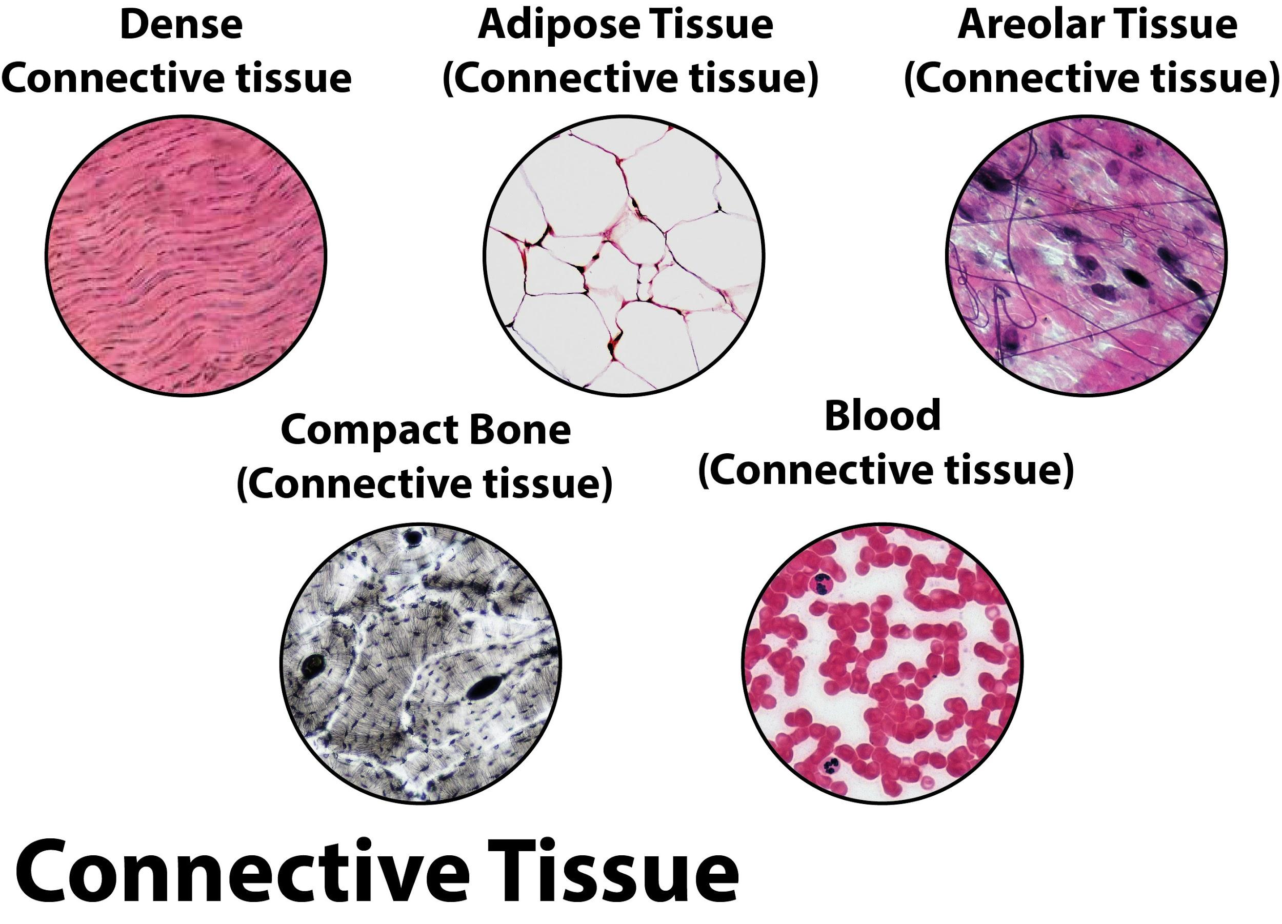
What is fibromyxoid stroma? This term, often encountered in pathology reports, refers to a specific type of connective tissue characterized by a unique blend of fibrous and myxoid components. Found in various tumors and conditions, fibromyxoid stroma plays a significant role in both diagnosis and prognosis, offering insights into the nature and behavior of the underlying disease.
Fibromyxoid stroma is a fascinating example of how microscopic structures can hold immense clinical significance. Its presence or absence, along with its specific features, can provide valuable information to clinicians and researchers, helping them understand the intricacies of disease processes and guide appropriate treatment strategies.
Definition of Fibromyxoid Stroma
:max_bytes(150000):strip_icc()/dense_irregular_conn_tissue-5b68c5db46e0fb0050b3a01b.jpg)
Fibromyxoid stroma is a type of connective tissue found in various tissues and organs, particularly in the context of pathology. It’s often associated with certain types of tumors, where its presence can have implications for diagnosis and treatment.
Components of Fibromyxoid Stroma, What is fibromyxoid stroma
Fibromyxoid stroma is characterized by a combination of fibrous connective tissue and myxoid material.
- Fibrous Connective Tissue: This component is composed of collagen fibers, which provide structural support and strength to the stroma. These fibers are typically arranged in a haphazard or loose pattern, contributing to the overall appearance of fibromyxoid stroma.
- Myxoid Material: Myxoid material is a gel-like substance rich in hyaluronic acid and other glycosaminoglycans. It gives the stroma its characteristic mucoid or gelatinous appearance. The presence of myxoid material often contributes to the soft, rubbery texture of fibromyxoid stroma.
Microscopic Appearance of Fibromyxoid Stroma
Under a microscope, fibromyxoid stroma typically appears as a pale, eosinophilic (pink-staining) area with a delicate, reticular (net-like) pattern. The collagen fibers are often thin and sparsely distributed, while the myxoid material creates a background of a slightly granular or amorphous appearance. The overall impression is of a loosely organized, somewhat translucent tissue.
Occurrence and Significance

Fibromyxoid stroma is a distinctive histological feature found in various tumors and conditions, playing a significant role in their classification, diagnosis, and prognosis. Its presence or absence can provide valuable insights into the nature and behavior of these diseases.
Types of Tumors and Conditions
The presence of fibromyxoid stroma is commonly observed in several types of tumors and conditions. Here’s a breakdown of some prominent examples:
- Fibromyxoid Sarcoma: This rare, aggressive soft tissue sarcoma is characterized by the presence of a prominent fibromyxoid stroma, contributing to its distinct histological appearance.
- Liposarcoma: Some subtypes of liposarcoma, particularly the myxoid liposarcoma, exhibit fibromyxoid stroma as a key characteristic.
- Mesenchymal Tumors: Fibromyxoid stroma can be found in a range of mesenchymal tumors, including fibromas, myxomas, and chondromas, highlighting its association with connective tissue elements.
- Desmoid Tumors: Also known as aggressive fibromatosis, desmoid tumors often display fibromyxoid stroma, contributing to their infiltrative growth pattern.
- Inflammatory Conditions: Fibromyxoid stroma can also occur in inflammatory conditions, such as nodular fasciitis, where it reflects the reactive nature of the tissue.
Clinical Significance
The presence of fibromyxoid stroma in various diseases holds significant clinical implications, influencing diagnostic and prognostic considerations. Here’s how:
- Diagnosis: The presence of fibromyxoid stroma can aid in differentiating between various tumor types, particularly in soft tissue sarcomas. For instance, the presence of fibromyxoid stroma in a soft tissue tumor strongly suggests a diagnosis of fibromyxoid sarcoma.
- Prognosis: In some cases, the presence or absence of fibromyxoid stroma can provide insights into the tumor’s aggressiveness and potential for recurrence. For example, in liposarcoma, the presence of myxoid stroma may indicate a more indolent behavior, while its absence might suggest a more aggressive variant.
- Treatment: The presence of fibromyxoid stroma can influence treatment strategies. For example, in fibromyxoid sarcoma, the tumor’s tendency for local recurrence often necessitates wide surgical margins to achieve adequate control.
Implications in Diagnosis and Prognosis
The presence or absence of fibromyxoid stroma can significantly impact the diagnosis and prognosis of various diseases.
- Presence: The presence of fibromyxoid stroma can be a crucial diagnostic feature, particularly in tumors where its presence is characteristic, such as fibromyxoid sarcoma. Additionally, it can indicate a specific subtype of a tumor, as seen in myxoid liposarcoma. Furthermore, in some cases, the presence of fibromyxoid stroma can be associated with a more aggressive tumor behavior, requiring more intensive treatment strategies.
- Absence: The absence of fibromyxoid stroma can also be informative. In some cases, its absence may rule out certain diagnoses, such as fibromyxoid sarcoma. Additionally, its absence might indicate a less aggressive tumor variant, potentially leading to a more favorable prognosis.
Cellular Components
Fibromyxoid stroma is a specialized type of connective tissue that is characterized by its unique cellular composition. It is a diverse environment containing a variety of cells that contribute to its formation, function, and potential for both benign and malignant growth.
Types of Cells
The cellular composition of fibromyxoid stroma is diverse, reflecting its complex role in tissue development and function. Here are some of the major cell types found within fibromyxoid stroma:
- Fibroblasts: These are the primary cells responsible for synthesizing and depositing the extracellular matrix (ECM) components of fibromyxoid stroma. They produce collagen fibers, proteoglycans, and other structural components that contribute to the stroma’s unique characteristics.
- Myofibroblasts: These cells share features of both fibroblasts and smooth muscle cells. They possess contractile properties, allowing them to regulate the tension and organization of the stroma. Myofibroblasts play a crucial role in wound healing and tissue remodeling.
- Mesenchymal Stem Cells: These undifferentiated cells have the potential to differentiate into various cell types, including fibroblasts, myofibroblasts, and other stromal cells. They contribute to the dynamic nature of fibromyxoid stroma and its ability to respond to various stimuli.
- Immune Cells: Fibromyxoid stroma can also contain various immune cells, including macrophages, lymphocytes, and mast cells. These cells play a role in immune surveillance and inflammation, influencing the stroma’s response to injury, infection, and tumor growth.
- Endothelial Cells: These cells line the blood vessels within the stroma, facilitating nutrient and oxygen delivery, as well as waste removal. They contribute to the vascularization of the stroma and its ability to support tissue growth and repair.
Cellular Atypia and Malignancy
While fibromyxoid stroma is generally benign, there is potential for cellular atypia and malignancy within this tissue. This is especially relevant in the context of certain tumors, such as fibromyxoid sarcoma.
- Cellular Atypia: This refers to abnormal changes in the appearance of cells within the stroma. It can involve variations in cell size, shape, and nuclear features. Cellular atypia is often a sign of precancerous changes or early malignancy.
- Malignancy: In some cases, cells within fibromyxoid stroma can undergo malignant transformation, leading to the development of fibromyxoid sarcoma. This type of cancer is characterized by uncontrolled growth and spread of atypical cells within the stroma.
The presence of cellular atypia or malignancy in fibromyxoid stroma is often associated with specific genetic alterations or environmental factors. These changes can disrupt the normal balance of cell growth and differentiation, leading to the formation of tumors.
Molecular Basis

The molecular basis of fibromyxoid stroma is a complex interplay of signaling pathways, gene expression, and protein interactions. Understanding these mechanisms is crucial for developing targeted therapies for fibromyxoid stroma-associated diseases.
Key Molecular Pathways and Signaling Molecules
Several key molecular pathways and signaling molecules are involved in the development and regulation of fibromyxoid stroma. These pathways are responsible for the production of extracellular matrix components, cell migration, and angiogenesis, all of which contribute to the formation and maintenance of fibromyxoid stroma.
- Transforming Growth Factor-beta (TGF-β) Pathway: TGF-β is a potent stimulator of fibromyxoid stroma formation. It activates downstream signaling pathways, leading to increased production of extracellular matrix components like collagen and fibronectin. TGF-β also promotes the differentiation of fibroblasts into myofibroblasts, which are responsible for the contractile nature of fibromyxoid stroma.
- Platelet-Derived Growth Factor (PDGF) Pathway: PDGF plays a critical role in the recruitment and proliferation of fibroblasts, which are essential for fibromyxoid stroma formation. PDGF signaling also stimulates the production of extracellular matrix components.
- Wnt Signaling Pathway: Wnt signaling is involved in the regulation of cell proliferation, differentiation, and migration. In the context of fibromyxoid stroma, Wnt signaling can contribute to the expansion of fibroblasts and the formation of a dense extracellular matrix.
- Hedgehog Signaling Pathway: The Hedgehog pathway is involved in the development and maintenance of tissues. Aberrant Hedgehog signaling has been implicated in the development of fibromyxoid stroma in certain diseases.
Role of Specific Genes and Proteins
Specific genes and proteins play crucial roles in the formation and maintenance of fibromyxoid stroma. These molecules can be targets for therapeutic intervention.
- Collagen genes (COL1A1, COL1A2, COL3A1): These genes encode the major components of collagen, a fibrous protein that forms the structural framework of fibromyxoid stroma. Increased expression of these genes leads to increased collagen deposition, contributing to the dense, fibrous nature of fibromyxoid stroma.
- Fibronectin (FN1): Fibronectin is an adhesive protein that helps cells attach to the extracellular matrix. Elevated levels of fibronectin contribute to the adhesion and migration of cells within fibromyxoid stroma.
- Matrix Metalloproteinases (MMPs): MMPs are enzymes that degrade the extracellular matrix. MMPs are involved in the remodeling of fibromyxoid stroma, and their dysregulation can contribute to the progression of diseases associated with fibromyxoid stroma.
- α-Smooth Muscle Actin (α-SMA): α-SMA is a marker of myofibroblasts, which are contractile cells that contribute to the rigidity and contractility of fibromyxoid stroma.
Potential for Targeted Therapies
The molecular understanding of fibromyxoid stroma has opened avenues for developing targeted therapies.
- TGF-β inhibitors: Blocking TGF-β signaling could reduce the production of extracellular matrix components and the differentiation of fibroblasts into myofibroblasts, thereby inhibiting the formation and progression of fibromyxoid stroma.
- PDGF inhibitors: Targeting PDGF signaling could reduce the recruitment and proliferation of fibroblasts, thereby limiting the expansion of fibromyxoid stroma.
- MMP inhibitors: MMP inhibitors could modulate the remodeling of fibromyxoid stroma, potentially preventing the progression of diseases associated with fibromyxoid stroma.
- Gene therapy: Gene therapy approaches could be used to target specific genes involved in fibromyxoid stroma formation, such as collagen genes or α-SMA.
Diagnostic Considerations
Fibromyxoid stroma is a distinctive type of connective tissue that can be identified through histopathological examination. The distinctive features of fibromyxoid stroma, including its unique cellular composition and extracellular matrix, are crucial for its accurate diagnosis.
Histopathological Examination
Histopathological examination plays a central role in the diagnosis of fibromyxoid stroma. A pathologist examines tissue samples under a microscope to identify the characteristic features of fibromyxoid stroma. These features include the presence of spindle-shaped fibroblasts, a myxoid matrix, and a loose, interwoven arrangement of collagen fibers. The pathologist also looks for other features, such as the presence of inflammatory cells or blood vessels, which can help to differentiate fibromyxoid stroma from other types of connective tissue.
Distinguishing Features of Fibromyxoid Stroma
Fibromyxoid stroma is characterized by a unique combination of features that set it apart from other types of connective tissue. These features include:
- Spindle-shaped fibroblasts: Fibromyxoid stroma contains spindle-shaped fibroblasts, which are elongated cells with a central nucleus and a cytoplasm that is often eosinophilic.
- Myxoid matrix: The extracellular matrix of fibromyxoid stroma is characterized by a myxoid appearance, which is a result of the high content of hyaluronic acid and other glycosaminoglycans. This matrix is often described as “gelatinous” or “mucoid”.
- Loose, interwoven collagen fibers: The collagen fibers in fibromyxoid stroma are typically arranged in a loose, interwoven pattern. These fibers are often thin and delicate, and they may be stained with Masson’s trichrome stain.
- Low cellularity: Fibromyxoid stroma is generally characterized by a low cellularity, with a relatively small number of cells compared to the amount of extracellular matrix.
Potential for Misdiagnosis
While histopathological examination is essential for the accurate diagnosis of fibromyxoid stroma, it is important to be aware of the potential for misdiagnosis. Fibromyxoid stroma can be mistaken for other types of connective tissue, such as myxoid liposarcoma or myxofibrosarcoma. Careful evaluation of the tissue sample, including the presence of other features, such as the presence of atypical cells or mitotic figures, is crucial to avoid misdiagnosis.
Clinical Management
The clinical management of tumors and conditions associated with fibromyxoid stroma is complex and often requires a multidisciplinary approach involving surgeons, pathologists, oncologists, and radiologists. Treatment strategies are tailored to the specific type of tumor, its stage, and the patient’s overall health.
Surgical Management
Surgery is the primary treatment modality for most tumors with fibromyxoid stroma. The goal of surgery is to completely remove the tumor, which can be challenging due to the infiltrative nature of these tumors. In some cases, the tumor may be located in a difficult-to-access area, requiring complex surgical techniques.
Radiation Therapy
Radiation therapy is often used as an adjunct to surgery, particularly for high-risk tumors or those that cannot be completely removed. Radiation therapy uses high-energy rays to kill cancer cells and shrink tumors.
Chemotherapy
Chemotherapy is less commonly used in the treatment of tumors with fibromyxoid stroma. It is often reserved for advanced or metastatic disease, and its effectiveness can be limited.
Ongoing Research and Future Advancements
Ongoing research is focused on understanding the molecular basis of fibromyxoid stroma and developing targeted therapies. For example, researchers are investigating the role of specific genes and proteins in tumor development and growth, which may lead to the development of new drugs that can specifically target these pathways.
Understanding fibromyxoid stroma is essential for anyone involved in the diagnosis and treatment of a wide range of conditions. From its unique microscopic appearance to its potential clinical implications, this connective tissue serves as a key indicator of disease progression and response to therapy. Continued research into the molecular basis of fibromyxoid stroma promises to unlock even greater insights into its role in human health and disease, ultimately leading to improved patient care.
User Queries: What Is Fibromyxoid Stroma
What are the most common tumors where fibromyxoid stroma is found?
Fibromyxoid stroma is commonly found in soft tissue tumors, including fibrosarcoma, myxofibrosarcoma, and low-grade fibromyxoid sarcoma.
Can fibromyxoid stroma be seen with the naked eye?
No, fibromyxoid stroma is typically microscopic and requires specialized staining techniques to be visualized under a microscope.
What is the difference between fibromyxoid stroma and myxoid stroma?
Fibromyxoid stroma contains both fibrous and myxoid components, while myxoid stroma is primarily composed of myxoid material. The presence of fibrous elements distinguishes fibromyxoid stroma from myxoid stroma.
Is fibromyxoid stroma always associated with cancer?
No, fibromyxoid stroma can be found in both benign and malignant conditions. However, its presence in certain tumors can be indicative of aggressive behavior.


