What is stroma reticular fibers – What are stroma reticular fibers? Think of them as the invisible scaffolding that holds your body together, a delicate network of fibers woven throughout many of your tissues. These fibers, made of a special type of protein called collagen, provide structure and support, and they play a critical role in the development and function of many vital organs.
From the filtering system of your lymph nodes to the blood-producing factory of your bone marrow, reticular fibers are everywhere, quietly doing their important job.
While they may not be as glamorous as muscles or as complex as neurons, reticular fibers are essential for maintaining the integrity and health of your tissues. They provide a framework for cells to grow and interact, and they help to regulate the flow of fluids and nutrients. But their story doesn’t end there. Reticular fibers also play a critical role in wound healing and tissue regeneration, and they are implicated in a variety of diseases, from cancer to fibrosis.
Introduction to Stroma: What Is Stroma Reticular Fibers
Stroma, derived from the Greek word “στρῶμα” (strōma) meaning “bed,” refers to the supporting framework or matrix of an organ or tissue. It acts as the structural foundation, providing a scaffold for the functional cells and tissues to reside and operate. Stroma is often composed of various components, including connective tissue, extracellular matrix, and sometimes specialized cells, all working in concert to maintain the integrity and function of the tissue.
Types of Tissues with Stroma
Stroma is ubiquitous in biological tissues, playing a crucial role in supporting and organizing various cell types. Here are some examples of tissues where stroma is found:
- Connective Tissues: Stroma is the primary component of connective tissues, such as cartilage, bone, and blood. It provides structural support, elasticity, and tensile strength, allowing these tissues to withstand stress and strain.
- Epithelial Tissues: Stroma supports epithelial tissues, which line the surfaces of organs and cavities. It provides a base for epithelial cells to attach to, ensuring their proper arrangement and function.
- Muscle Tissues: Stroma in muscle tissues provides structural support and allows for the efficient transmission of force during contraction. It also facilitates the vascularization and innervation of muscle fibers.
- Nervous Tissues: Stroma in nervous tissues provides a framework for the intricate network of neurons and glial cells. It supports the delicate nerve fibers, ensuring proper signal transmission and integration.
- Glands: Stroma in glands provides support for the secretory cells and ducts, facilitating the production and release of hormones and other substances.
Functions of Stroma
The functions of stroma are multifaceted and essential for maintaining the integrity and functionality of tissues and organs.
- Structural Support: Stroma provides a framework that holds cells and tissues together, preventing them from dispersing and ensuring their proper organization.
- Nutrient and Waste Exchange: Stroma facilitates the diffusion of nutrients, oxygen, and other essential molecules to the cells within the tissue. It also allows for the removal of waste products from the cells.
- Cell Communication: Stroma plays a crucial role in cell signaling and communication, providing a pathway for the exchange of chemical and physical signals between cells.
- Tissue Repair and Regeneration: Stroma is involved in tissue repair and regeneration processes, providing a scaffold for new cells to grow and differentiate.
- Immune Response: Stroma can participate in immune responses by providing a platform for immune cells to interact and migrate within the tissue.
Reticular Fibers

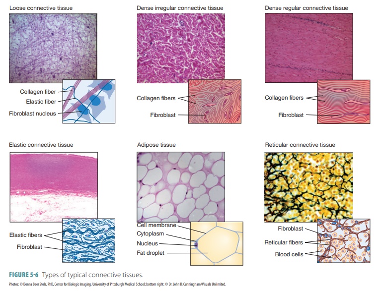
Reticular fibers are a type of collagen fiber found in various tissues throughout the body. They are known for their delicate, interwoven network structure, providing support and framework for cells and tissues.
Structure and Composition
Reticular fibers are composed primarily of type III collagen, a protein that forms thin, branching fibers. These fibers are coated with a glycoprotein called reticulin, which enhances their ability to bind with other cells and tissues. The presence of reticulin, along with the thin and branching nature of type III collagen, contributes to the unique characteristics of reticular fibers.
- Reticular fibers are thinner and more delicate than collagen fibers, forming a fine mesh-like network.
- They are typically found in tissues that require flexibility and support, such as the stroma of lymphoid organs (e.g., spleen, lymph nodes), bone marrow, liver, and smooth muscle.
- The delicate nature of reticular fibers allows for the passage of cells and fluids, while providing structural support.
Differences from Collagen Fibers
Reticular fibers differ from collagen fibers in several key aspects:
- Composition: Reticular fibers are composed primarily of type III collagen, while collagen fibers are predominantly made of type I collagen.
- Thickness: Reticular fibers are thinner and more delicate than collagen fibers.
- Branching: Reticular fibers are highly branched and interwoven, forming a fine network, whereas collagen fibers are typically thicker and less branched.
- Location: Reticular fibers are commonly found in soft tissues, while collagen fibers are more abundant in connective tissues, such as tendons and ligaments.
- Function: Reticular fibers provide a delicate framework for cells and tissues, while collagen fibers offer strength and support.
Formation and Assembly of Reticular Fibers

Reticular fibers, a type of collagen fiber, are synthesized and assembled through a complex process involving specialized cells and molecular interactions. This process is crucial for the formation of a supportive framework within various tissues and organs, particularly in the lymphatic system and hematopoietic tissues.
Role of Reticular Cells in Fiber Formation
Reticular cells, also known as fibroblasts, are responsible for the synthesis and secretion of the protein precursors that make up reticular fibers. These cells are found in various tissues and play a vital role in maintaining the structural integrity of the extracellular matrix.
- Synthesis of Procollagen III: Reticular cells synthesize procollagen type III, the precursor protein of reticular fibers. This process involves the translation of genetic information from DNA into mRNA, which then directs the synthesis of procollagen III molecules within the cell.
- Assembly of Procollagen Molecules: Procollagen III molecules assemble into triple helix structures, forming long, fibrillar strands. These strands are then secreted from the reticular cells into the extracellular matrix.
- Cleavage and Cross-linking: Once outside the cell, specific enzymes cleave the terminal propeptides from the procollagen III molecules. This cleavage step exposes the collagen molecules, allowing them to self-assemble into a network of fibers.
- Formation of Reticular Fiber Network: The assembled collagen molecules interact with other components of the extracellular matrix, including glycosaminoglycans and proteoglycans. These interactions contribute to the formation of a three-dimensional network of reticular fibers, providing structural support and creating a framework for cells and tissues.
Interaction with Other Extracellular Matrix Components
Reticular fibers interact with other components of the extracellular matrix, forming a complex and dynamic network that supports and organizes cells and tissues. This interaction is crucial for maintaining tissue integrity and regulating cell function.
- Glycosaminoglycans: Reticular fibers interact with glycosaminoglycans, negatively charged polysaccharides that attract water and contribute to the hydration and elasticity of the extracellular matrix. These interactions help maintain the spacing between fibers and provide structural support.
- Proteoglycans: Reticular fibers also interact with proteoglycans, large molecules composed of a core protein attached to glycosaminoglycans. Proteoglycans bind to collagen fibers and other components of the extracellular matrix, forming a complex network that regulates cell adhesion, migration, and signaling.
- Other Collagen Types: Reticular fibers can also interact with other collagen types, such as type I collagen, forming a more robust and organized network. This interaction contributes to the strength and resilience of tissues, particularly in areas that experience mechanical stress.
Clinical Significance of Reticular Fibers
Reticular fibers, despite their delicate structure, play a crucial role in various physiological processes, particularly in tissue repair and regeneration. Their unique properties and interactions with other components of the extracellular matrix contribute to the overall health and function of tissues. However, alterations in their structure and organization can lead to disease development, highlighting their clinical significance.
Role in Tissue Repair and Regeneration
Reticular fibers act as a scaffold during tissue repair and regeneration. They provide a framework for the migration and proliferation of cells involved in the healing process, such as fibroblasts, endothelial cells, and immune cells. This scaffolding action is essential for the formation of new tissue and the restoration of tissue function.
- During wound healing, reticular fibers provide a structural support for the ingrowth of new blood vessels, which are essential for delivering oxygen and nutrients to the healing tissue.
- Reticular fibers also facilitate the migration and proliferation of fibroblasts, which produce collagen, a key component of the extracellular matrix, and contribute to the formation of scar tissue.
Alterations in Reticular Fiber Structure and Disease Development
Disruptions in the structure and organization of reticular fibers can contribute to the development of various diseases. These disruptions can occur due to genetic mutations, environmental factors, or aging.
- Fibrosis: Excessive deposition of collagen and other extracellular matrix components, including reticular fibers, can lead to fibrosis, a condition characterized by the thickening and scarring of tissues. This can occur in various organs, including the liver, lungs, and kidneys, impairing their function.
- Cancer: Alterations in reticular fiber structure can promote tumor growth and metastasis. Reticular fibers can provide a framework for tumor cells to invade surrounding tissues and spread to distant sites.
- Immune Disorders: Reticular fibers can be involved in immune responses. Aberrant reticular fiber organization can contribute to autoimmune diseases, such as rheumatoid arthritis, where the immune system attacks the body’s own tissues.
Examples of Diseases Associated with Reticular Fiber Abnormalities, What is stroma reticular fibers
Several diseases are associated with abnormalities in reticular fiber structure. These abnormalities can involve alterations in the quantity, distribution, or composition of reticular fibers.
- Scleroderma: This autoimmune disease is characterized by excessive collagen deposition in the skin and internal organs, leading to thickening and hardening of the tissues. This excessive collagen deposition can involve reticular fibers, contributing to the characteristic fibrosis observed in scleroderma.
- Cirrhosis: This chronic liver disease is characterized by the formation of scar tissue in the liver, leading to impaired liver function. Reticular fibers play a role in the development of cirrhosis by providing a scaffold for the deposition of collagen and other extracellular matrix components.
- Pulmonary Fibrosis: This lung disease is characterized by scarring and thickening of the lung tissue, leading to difficulty breathing. Reticular fibers are involved in the development of pulmonary fibrosis by providing a framework for the deposition of collagen and other extracellular matrix components.
So next time you think about the complex workings of your body, remember the tiny but mighty reticular fibers. They are the unsung heroes of your health, quietly working behind the scenes to keep everything running smoothly. From their role in tissue development to their involvement in disease, reticular fibers are a fascinating and important part of your biological story.
FAQ Explained
What is the difference between reticular fibers and collagen fibers?
Reticular fibers are a specialized type of collagen fiber that forms delicate networks. They are thinner and more branched than other types of collagen fibers, and they are typically found in tissues that require flexibility and support, like the lymphatic system and bone marrow.
What are some diseases associated with reticular fiber abnormalities?
Alterations in reticular fiber structure can contribute to a variety of diseases, including fibrosis, cancer, and autoimmune disorders. For example, in fibrosis, the overproduction of reticular fibers can lead to scarring and tissue damage. In cancer, reticular fibers can be altered to promote tumor growth and spread.


